Dear Customer, If you experience any text readability or display issues on our website, please update your web browser to the latest version
.
See More
Dear Customer, If you experience any text readability or display issues on our website, please update your web browser to the latest version
.
See MoreDear Customer, If you experience any text readability or display issues on our website, please update your web browser to the latest version
.
This page has been recently translated and is available in French now.
Looks like you're visiting us from {countryName}.
Would you like to stay on the current location site or be switched to your location?
Flow cytometric analysis of CD4 expression on human peripheral blood lymphocytes. Human whole blood was stained at 4°C with the BD Horizon™ BV421 Mouse anti-Human CD4 antibody (Cat. No. 562424/562425; solid line histogram) or with a BD Horizon™ BV421 Mouse IgG1, κ Isotype Control (Cat. No. 562438; dashed line histogram). The erythrocytes were lysed with BD Pharm Lyse™ Lysing Buffer (Cat. No. 555899). The fluorescence histograms were derived from events with the forward and side light-scatter characteristics of viable lymphocytes. Flow cytometry was performed using a BD FACSCanto™ II Flow Cytometer System.
Immunohistofluorescent analysis of CD4 expression by cells within human tonsil. A human tonsil cryosection (5 µm) was fixed with BD Cytofix™ Fixation Buffer (Cat. No. 554655), blocked with 5% goat serum and 1% BSA diluted in 1x PBS, and stained with BD Pharmingen™ Purified Mouse Anti-Human CD8 antibody (Cat. No. 555364) followed by BD Horizon™ BV480 Goat Anti-Mouse Ig second step antibody (Cat. No. 564877, pseudo-colored green). Sections were thoroughly washed, then stained with BD Horizon™ BV421 Mouse Anti-Human CD4 antibody (Cat. No. 562424/562425, pseudo-colored red) and Alexa Fluor® 488 Mouse Anti-Human CD19 antibody (Cat. No. 557697, pseudo-colored blue). Images were captured on a standard epifluorescence microscope. Original magnification, 20x.
Flow cytometric analysis of CD4 expression on human peripheral blood lymphocytes. Human whole blood was stained at 4°C with the BD Horizon™ BV421 Mouse anti-Human CD4 antibody (Cat. No. 562424/562425; solid line histogram) or with a BD Horizon™ BV421 Mouse IgG1, κ Isotype Control (Cat. No. 562438; dashed line histogram). The erythrocytes were lysed with BD Pharm Lyse™ Lysing Buffer (Cat. No. 555899). The fluorescence histograms were derived from events with the forward and side light-scatter characteristics of viable lymphocytes. Flow cytometry was performed using a BD FACSCanto™ II Flow Cytometer System.
Immunohistofluorescent analysis of CD4 expression by cells within human tonsil. A human tonsil cryosection (5 µm) was fixed with BD Cytofix™ Fixation Buffer (Cat. No. 554655), blocked with 5% goat serum and 1% BSA diluted in 1x PBS, and stained with BD Pharmingen™ Purified Mouse Anti-Human CD8 antibody (Cat. No. 555364) followed by BD Horizon™ BV480 Goat Anti-Mouse Ig second step antibody (Cat. No. 564877, pseudo-colored green). Sections were thoroughly washed, then stained with BD Horizon™ BV421 Mouse Anti-Human CD4 antibody (Cat. No. 562424/562425, pseudo-colored red) and Alexa Fluor® 488 Mouse Anti-Human CD19 antibody (Cat. No. 557697, pseudo-colored blue). Images were captured on a standard epifluorescence microscope. Original magnification, 20x.
Flow cytometric analysis of CD4 expression on human peripheral blood lymphocytes. Human whole blood was stained at 4°C with the BD Horizon™ BV421 Mouse anti-Human CD4 antibody (Cat. No. 562424/562425; solid line histogram) or with a BD Horizon™ BV421 Mouse IgG1, κ Isotype Control (Cat. No. 562438; dashed line histogram). The erythrocytes were lysed with BD Pharm Lyse™ Lysing Buffer (Cat. No. 555899). The fluorescence histograms were derived from events with the forward and side light-scatter characteristics of viable lymphocytes. Flow cytometry was performed using a BD FACSCanto™ II Flow Cytometer System.
Immunohistofluorescent analysis of CD4 expression by cells within human tonsil. A human tonsil cryosection (5 µm) was fixed with BD Cytofix™ Fixation Buffer (Cat. No. 554655), blocked with 5% goat serum and 1% BSA diluted in 1x PBS, and stained with BD Pharmingen™ Purified Mouse Anti-Human CD8 antibody (Cat. No. 555364) followed by BD Horizon™ BV480 Goat Anti-Mouse Ig second step antibody (Cat. No. 564877, pseudo-colored green). Sections were thoroughly washed, then stained with BD Horizon™ BV421 Mouse Anti-Human CD4 antibody (Cat. No. 562424/562425, pseudo-colored red) and Alexa Fluor® 488 Mouse Anti-Human CD19 antibody (Cat. No. 557697, pseudo-colored blue). Images were captured on a standard epifluorescence microscope. Original magnification, 20x.
Any use of products other than the permitted use without the express written authorization of Becton, Dickinson and Company is strictly prohibited.
For optimal and reproducible results, BD Horizon Brilliant Stain Buffer should be used anytime two or more BD Horizon Brilliant dyes are used in the same experiment. Fluorescent dye interactions may cause staining artifacts which may affect data interpretation. The BD Horizon Brilliant Stain Buffer was designed to minimize these interactions. More information can be found in the Technical Data Sheet of the BD Horizon Brilliant Stain Buffer (Cat. No. 563794).
The RPA-T4 monoclonal antibody specifically binds to CD4, a 59 kDa single-chain transmembrane glycoprotein that is expressed on T-helper/inducer cell populations. CD4 is also expressed on thymocyte subsets and at lower levels on monocytes and macrophages. CD4 functions as a co-receptor in MHC class II-restricted antigen-induced T cell activation and as a receptor for human immunodeficiency viruses (HIV). This antibody binds to the D1 domain (CDR1 and CDR3 epitopes) of the CD4 antigen and reacts with approximately 80% of thymocytes and 45% of peripheral blood lymphocytes. RPA-T4 is capable of blocking HIV-1, gp120, and inhibits syncytium formation.
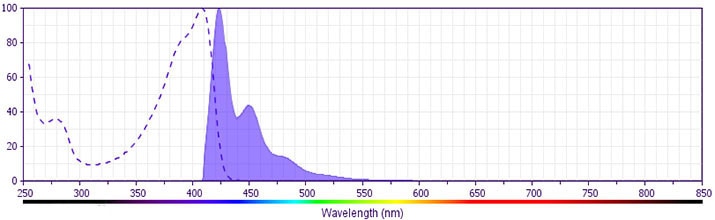
The antibody was conjugated to BD Horizon™ BV421 which is part of the BD Horizon Brilliant™ Violet family of dyes. With an Ex Max of 407-nm and Em Max at 421-nm, BD Horizon™ BV421 can be excited by the violet laser and detected in the standard Pacific Blue™ filter set (eg, 450/50-nm filter). BD Horizon™ BV421 conjugates are very bright, often exhibiting a 10 fold improvement in brightness compared to Pacific Blue™ conjugates.
Please refer to Support Documents for Quality Certificates
Global - Refer to manufacturer's instructions for use and related User Manuals and Technical data sheets before using this products as described
Comparisons, where applicable, are made against older BD Technology, manual methods or are general performance claims. Comparisons are not made against non-BD technologies, unless otherwise noted.
For Research Use Only. Not for use in diagnostic or therapeutic procedures.