







Preparation And Storage


Recommended Assay Procedures
BD™ CompBeads can be used as surrogates to assess fluorescence spillover (Compensation). When fluorochrome conjugated antibodies are bound to CompBeads, they have spectral properties very similar to cells. However, for some fluorochromes there can be small differences in spectral emissions compared to cells, resulting in spillover values that differ when compared to biological controls. It is strongly recommended that when using a reagent for the first time, users compare the spillover on cell and CompBead to ensure that BD Comp beads are appropriate for your specific cellular application.
Product Notices
- This reagent has been pre-diluted for use at the recommended Volume per Test. We typically use 1 × 10^6 cells in a 100-µl experimental sample (a test).
- An isotype control should be used at the same concentration as the antibody of interest.
- Source of all serum proteins is from USDA inspected abattoirs located in the United States.
- Caution: Sodium azide yields highly toxic hydrazoic acid under acidic conditions. Dilute azide compounds in running water before discarding to avoid accumulation of potentially explosive deposits in plumbing.
- Please observe the following precautions: Absorption of visible light can significantly alter the energy transfer occurring in any tandem fluorochrome conjugate; therefore, we recommend that special precautions be taken (such as wrapping vials, tubes, or racks in aluminum foil) to prevent exposure of conjugated reagents, including cells stained with those reagents, to room illumination.
- For fluorochrome spectra and suitable instrument settings, please refer to our Multicolor Flow Cytometry web page at www.bdbiosciences.com/colors.
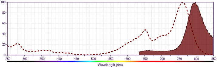
- BD APC-H7 is a tandem conjugate and an analog of APC-Cy7 with the same spectral properties. It has decreased intensity but it is engineered for greater stability and less spillover in the APC channel and consequently offers better performance than APC-Cy7. It has an absorption maximum of approximately 650 nm. When excited by light from a red laser, the APC fluorochrome can transfer energy to the cyanine dye, which then emits at a longer wavelength. The resulting fluorescent emission maximum is approximately 767 nm. BD recommends that a 750-nm longpass filter be used along with a red-sensitive detector such as the Hamamatsu R3896 PMT. As with APC-Cy7 special filters are required when using APC-H7 in conjunction with APC. Note: Although our APC-H7 products demonstrate higher lot-to lot consistency than other APC tandem conjugate products, and every effort is made to minimize the lot-to-lot variation in residual emission from APC, it is strongly recommended that every lot be tested for differences in the amount of compensation required and that individual compensation controls are run for each APC-H7 conjugate.
- Although BD APC-H7 is engineered to minimize spillover to the APC channel and is more stable and less affected by light, temperature, and formaldehyde-based fixatives, compared to other APC-cyanine tandem dyes, it is still good practice to minimize as much as possible, any light, temperature and fixative exposure when working with all fluorescent conjugates.
- Species cross-reactivity detected in product development may not have been confirmed on every format and/or application.
- Please refer to http://regdocs.bd.com to access safety data sheets (SDS).
- Cy is a trademark of Global Life Sciences Solutions Germany GmbH or an affiliate doing business as Cytiva.
- Please refer to www.bdbiosciences.com/us/s/resources for technical protocols.
Companion Products



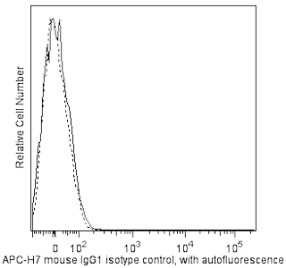
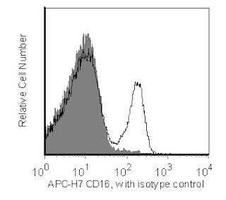
The 3G8 monoclonal antibody specifically recognizes CD16a and CD16b, low affinity receptors for the Fc region of IgG. CD16a is ~50-65 kDa type I transmembrane glycoprotein that is encoded by FCGR3A (Fc fragment of IgG receptor IIIa) which belongs to the immunoglobulin superfamily. CD16a is also known as Fc-gamma RIII-alpha (Fc-gamma RIIIa or FcγRIIIA) or FcRIIIa and is expressed on natural killer cells, activated monocytes, macrophages, γδ T cells, immature thymocytes, and mast cells. CD16a binds immune-complexed or aggregated IgG and associates with CD247/TCRζ in NK cells and FcεRIγ chains in phagocytes and mast cells to transduce intracellular signals. CD16a functions in antibody-dependent cellular cytotoxicity (ADCC) and other antibody-dependent responses including phagocytosis, cytokine production or mediator release. CD16b is a ~48 kDa glycophosyl-phosphatidylinositol (GPI)-linked form that is encoded by FCGR3B (Fc fragment of IgG receptor IIIb). CD16b is also known as Fc-gamma RIII-beta (Fc-gamma RIIIb or FcγRIIIB) or FcRIIIb and is expressed on neutrophils and activated eosinophils. The extracellular region of CD16b is highly homologous to CD16a. CD16b also serves as a receptor for the Fc region of IgG and can bind immune-complexed or aggregated IgG and may be involved in neutrophil adhesion.
The 3G8 antibody also crossreacts with a subset of peripheral blood lymphocytes and monocytes, but not granulocytes, of baboon, rhesus, and cynomolgus monkeys. Multicolor analysis reveals that the distribution on lymphocytes is similar to that found in human studies with the majority of CD16-positive lymphocytes being both CD3 and CD20 negative.
Development References (10)
-
Barclay NA, Brown MH, Birkeland ML, et al, ed. The Leukocyte Antigen FactsBook. San Diego, CA: Academic Press; 1997.
-
Fleit HB, Wright SD, Durie CJ, Valinsky JE, Unkeless JC. Ontogeny of Fc receptors and complement receptor (CR3) during human myeloid differentiation. J Clin Invest. 1984; 73(2):516-525. (Clone-specific: Flow cytometry, Fluorescence microscopy, Immunofluorescence, Immunoprecipitation, Radioimmunoassay). View Reference
-
Fleit HB, Wright SD, Unkeless JC. Human neutrophil Fc gamma receptor distribution and structure. Proc Natl Acad Sci U S A. 1982; 79(10):3275-3279. (Biology). View Reference
-
Knapp W. W. Knapp .. et al., ed. Leucocyte typing IV : white cell differentiation antigens. Oxford New York: Oxford University Press; 1989:1-1182.
-
Perussia B, Trinchieri G, Jackson A, et al. The Fc receptor for IgG on human natural killer cells: phenotypic, functional, and comparative studies with monoclonal antibodies. J Immunol. 1984; 133(1):180-189. (Clone-specific: Flow cytometry, Functional assay, Inhibition). View Reference
-
Schmidt RE. Non-lineage/natural killer section report: new and previously defined clusters. In: Knapp W. W. Knapp .. et al., ed. Leucocyte typing IV : white cell differentiation antigens. Oxford New York: Oxford University Press; 1989:517-542.
-
Stroncek DF, Skubitz KM, Plachta LB, et al. Alloimmune neonatal neutropenia due to an antibody to the neutrophil Fc-gamma receptor III with maternal deficiency of CD16 antigen. Blood. 1991; 77(7):1572-1580. (Biology). View Reference
-
Vossebeld PJ, Homburg CH, Roos D, Verhoeven AJ. The anti-Fc gamma RIII mAb 3G8 induces neutrophil activation via a cooperative actin of Fc gamma RIIIb and Fc gamma RIIa. Int J Biochem Cell Biol. 1997; 29(3):465-473. (Clone-specific). View Reference
-
Wirthmueller U, Kurosaki T, Murakami MS, Ravetch JV. Signal transduction by Fc gamma RIII (CD16) is mediated through the gamma chain. J Exp Med. 1992; 175(5):1381-1390. (Biology). View Reference
-
Zola H. Leukocyte and stromal cell molecules : the CD markers. Hoboken, N.J.: Wiley-Liss; 2007.
Please refer to Support Documents for Quality Certificates
Global - Refer to manufacturer's instructions for use and related User Manuals and Technical data sheets before using this products as described
Comparisons, where applicable, are made against older BD Technology, manual methods or are general performance claims. Comparisons are not made against non-BD technologies, unless otherwise noted.
For Research Use Only. Not for use in diagnostic or therapeutic procedures.
