Dear Customer, If you experience any text readability or display issues on our website, please update your web browser to the latest version
.
See More
Dear Customer, If you experience any text readability or display issues on our website, please update your web browser to the latest version
.
See MoreDear Customer, If you experience any text readability or display issues on our website, please update your web browser to the latest version
.
This page has been recently translated and is available in French now.
Looks like you're visiting us from {countryName}.
Would you like to stay on the current location site or be switched to your location?
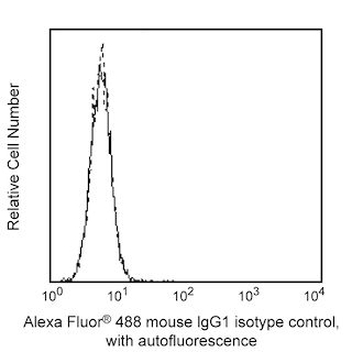
Analysis of TBK1 in transfected human epithelial cells. The 293 fetal kidney cell line (left panel) was either co-transfected with TBK1 and IRF-7 expression vectors (dashed histogram) or un-transfected (solid line). After 24 hours, the cells were fixed (BD Cytofix™ buffer, Cat. No. 554655) for 10 minutes at 37°C, then permeabilized (BD Phosflow™ Perm Buffer III, Cat. No. 558050) on ice for at least 30 minutes, and then stained with Alexa Fluor® 488 Mouse anti-TBK1 (Cat. No. 558697). Flow cytometry was performed on a BD FACSCalibur™ flow cytometry system. The specificity of mAb 637Ig11.2 was confirmed by western blot (right panel) using unconjugated antibody on lysates from untransfected (left panel) and TBK1/IRF-7 co-transfected (right panel) 293 cells. The TBK1-Myc tagged fusion protein is identified as a band of 84 kDa in the lysate of co-transfected cells.
Any use of products other than the permitted use without the express written authorization of Becton, Dickinson and Company is strictly prohibited.
NF-κB is a ubiquitously expressed transcription factor that regulates many cytokine and Ig genes. It is involved in immune, inflammatory, viral, and acute phase responses. In most cells, NF-κB is sequestered in an inactive cytoplasmic form via interactions with the inhibitory proteins IκBα, IκBβ, and IκBε. Stimulation induces the release, activation, and nuclear translocation of NF-κB. Release of NF-κB results from the phosphorylation and proteolytic degradation of the IκB proteins. Two cytokine-inducible IκB kinases (IKKα and IKKβ) phosphorylate and target the IκB proteins for degradation via the ubiquitin pathway. IKKγ/NEMO, a third member of the IKK complex, functions as a regulatory subunit and interacts directly with IKKβ. TBK1 (TANK-binding kinase 1, also known as T2K or NAK), a protein of 729 amino acids, is another member of the IKK family of kinases regulating NF-κB downstream of the tumor necrosis factor and Toll-like receptor pathways. TBK1 forms a complex with the adaptor proteins TANK (TRAF-associated NF-κB activator) and TRAF2 (TNF-receptor-associated factor 2), and this oligomer is required for activation and phosphorylation of TBK1 at serine 172.
The 637Ig11.2 monoclonal antibody recognizes human TBK1, regardless of phosphorylation status. Our in-house testing is performed on a cell line that has been co-transfected with TBK1 and IRF-7 because we have been unable to detect endogenous TBK1 by western blotting of lysates from activated cell lines (HeLa activated with PMA and A431 activated with PDGF). In the transfectants, the over-expression of TBK1 is necessary for phosphorylation of IRF-7, but the presence of IRF-7 does not affect TBK1 expression or phosphorylation. We confirmed that mAb 637Ig11.2 does not cross-react with IRF-7 by western blot analysis using the purified antibody.
Please refer to Support Documents for Quality Certificates
Global - Refer to manufacturer's instructions for use and related User Manuals and Technical data sheets before using this products as described
Comparisons, where applicable, are made against older BD Technology, manual methods or are general performance claims. Comparisons are not made against non-BD technologies, unless otherwise noted.
For Research Use Only. Not for use in diagnostic or therapeutic procedures.