-
Reagents
- Flow Cytometry Reagents
-
Western Blotting and Molecular Reagents
- Immunoassay Reagents
-
Single-Cell Multiomics Reagents
- BD® OMICS-Guard Sample Preservation Buffer
- BD® AbSeq Assay
- BD® Single-Cell Multiplexing Kit
- BD Rhapsody™ ATAC-Seq Assays
- BD Rhapsody™ Whole Transcriptome Analysis (WTA) Amplification Kit
- BD Rhapsody™ TCR/BCR Next Multiomic Assays
- BD Rhapsody™ Targeted mRNA Kits
- BD Rhapsody™ Accessory Kits
- BD® OMICS-One Protein Panels
-
Functional Assays
-
Microscopy and Imaging Reagents
-
Cell Preparation and Separation Reagents
-
- BD® OMICS-Guard Sample Preservation Buffer
- BD® AbSeq Assay
- BD® Single-Cell Multiplexing Kit
- BD Rhapsody™ ATAC-Seq Assays
- BD Rhapsody™ Whole Transcriptome Analysis (WTA) Amplification Kit
- BD Rhapsody™ TCR/BCR Next Multiomic Assays
- BD Rhapsody™ Targeted mRNA Kits
- BD Rhapsody™ Accessory Kits
- BD® OMICS-One Protein Panels
- Ireland (English)
-
Change country/language
Old Browser
This page has been recently translated and is available in French now.
Looks like you're visiting us from United States.
Would you like to stay on the current country site or be switched to your country?
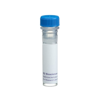
BD Transduction Laboratories™ Purified Mouse Anti-LAP2
Clone 27/LAP2 (RUO)


Western blot analysis of LAP2 on a RSV-3T3 cell lysate. Lane 1: 1:5000, lane 2: 1:10,000, lane 3: 1:20,000 dilution of the mouse anti-LAP2 antibody.

Immunofluorescence staining of HeLa cells (Human cervical epitheloid carcinoma; ATCC CCL-2.2).


BD Transduction Laboratories™ Purified Mouse Anti-LAP2

BD Transduction Laboratories™ Purified Mouse Anti-LAP2

Regulatory Status Legend
Any use of products other than the permitted use without the express written authorization of Becton, Dickinson and Company is strictly prohibited.
Preparation And Storage
Recommended Assay Procedures
Western blot: Please refer to http://www.bdbiosciences.com/pharmingen/protocols/Western_Blotting.shtml
Product Notices
- Since applications vary, each investigator should titrate the reagent to obtain optimal results.
- Please refer to www.bdbiosciences.com/us/s/resources for technical protocols.
- Caution: Sodium azide yields highly toxic hydrazoic acid under acidic conditions. Dilute azide compounds in running water before discarding to avoid accumulation of potentially explosive deposits in plumbing.
- Source of all serum proteins is from USDA inspected abattoirs located in the United States.
Data Sheets
Companion Products

.png?imwidth=320)
A specialized extension of the ER, the nuclear envelope (NE) forms the nuclear compartment boundary in eukaryotic cells. It contains numerous pore complexes and the nucleoplasmic side is linked to nuclear lamina. The nuclear lamina composes the structural framework for the NE and serves as a chromatin anchor site, thus, playing a major role in interphase nuclear organization. Many proteins are associated with lamina, particularly the LAPs (Lamina-Associated Polypeptides). LAP2 (also known as LAP2β) is a hydrophilic protein with a single transmembrane segment near the C-terminus. Thus, it has been defined as a type II integral membrane protein with the majority of its structure exposed to the nucleoplasm. LAP2 binding to lamins contributes to the attachment of the nuclear lamina to the inner nuclear membrane. LAP2 also binds to chromatin, implying its role in chromosomal organization during mitosis. Mitotic phosphorylation of LAP2 regulates its binding to lamins and chromosomes during the disassembly and reassembly of mitosis. Thus, LAP2 is a nuclear protein that plays a role in the organization of the NE during cell cycle progression.
Development References (5)
-
Dechat T, Gotzmann J, Stockinger A, et al. Detergent-salt resistance of LAP2alpha in interphase nuclei and phosphorylation-dependent association with chromosomes early in nuclear assembly implies functions in nuclear structure dynamics. EMBO J. 1998; 17(16):4887-4902. (Biology). View Reference
-
Furukawa K, Fritze CE, Gerace L. The major nuclear envelope targeting domain of LAP2 coincides with its lamin binding region but is distinct from its chromatin interaction domain. J Biol Chem. 1998; 273(7):4213-4219. (Biology). View Reference
-
Furukawa K, Pante N, Aebi U, Gerace L. Cloning of a cDNA for lamina-associated polypeptide 2 (LAP2) and identification of regions that specify targeting to the nuclear envelope. EMBO J. 1995; 14(8):1626-1636. (Biology). View Reference
-
Kimura T, Ito C, Watanabe S, et al. Mouse germ cell-less as an essential component for nuclear integrity. Mol Cell Biol. 2003; 23(4):1304-1315. (Biology: Immunofluorescence). View Reference
-
Rusan NM, Tulu US, Fagerstrom C, Wadsworth P. Reorganization of the microtubule array in prophase/prometaphase requires cytoplasmic dynein-dependent microtubule transport. J Biol Chem. 2002; 158(6):997-1003. (Biology: Immunofluorescence). View Reference
Please refer to Support Documents for Quality Certificates
Global - Refer to manufacturer's instructions for use and related User Manuals and Technical data sheets before using this products as described
Comparisons, where applicable, are made against older BD Technology, manual methods or are general performance claims. Comparisons are not made against non-BD technologies, unless otherwise noted.
For Research Use Only. Not for use in diagnostic or therapeutic procedures.