-
Reagents
- Flow Cytometry Reagents
-
Western Blotting and Molecular Reagents
- Immunoassay Reagents
-
Single-Cell Multiomics Reagents
- BD® OMICS-Guard Sample Preservation Buffer
- BD® AbSeq Assay
- BD® Single-Cell Multiplexing Kit
- BD Rhapsody™ ATAC-Seq Assays
- BD Rhapsody™ Whole Transcriptome Analysis (WTA) Amplification Kit
- BD Rhapsody™ TCR/BCR Next Multiomic Assays
- BD Rhapsody™ Targeted mRNA Kits
- BD Rhapsody™ Accessory Kits
- BD® OMICS-One Protein Panels
- BD OMICS-One™ WTA Next Assay
-
Functional Assays
-
Microscopy and Imaging Reagents
-
Cell Preparation and Separation Reagents
Old Browser
This page has been recently translated and is available in French now.
Looks like you're visiting us from {countryName}.
Would you like to stay on the current location site or be switched to your location?

Immunofluorescence
Fluorescence imaging–based techniques are highly useful for interrogating the structural and functional of aspects of cells and tissues. When combined with immunochemistry, their analytical power increases tremendously, enhancing their utility in a wide range of research and clinical applications, making them invaluable tools. Immunofluorescence microscopy revolutionized the field of cell biology by enabling live-cell imaging to visualize whole organelles. Immunohistochemistry and immunocytochemistry are common fluorescence-imaging techniques that combine the power of antigen-antibody binding for cell analysis.
What is immunofluorescence and what are its applications?
Immunofluorescence (IF) is a powerful immunostaining technique that utilizes microscopy to visualize fluorophore-conjugated antibodies bound to target proteins and other molecules of interest. IF is used to identify cell- and tissue-specific antigens in cells; visualize the presence or absence, cellular localization, and activation status of proteins; and analyze immune responses.
Principles and types of immunofluorescence
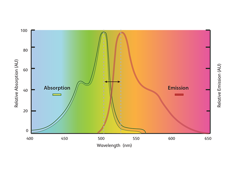
IF exploits the property of fluorescent molecules or fluorophores to absorb photons at a certain wavelength (absorption spectrum of the molecule) and emit them at a higher wavelength after a brief interval (emission spectrum) with an accompanied energy loss. The emitted fluorescence can be visualized by microscopy. Fluorophores can be excited by visible or UV light.
Fluorescent dyes with high photostability and fluorescence quantum yield are commercially available with excitation maxima spanning a range of wavelengths, from 400 to >700 nm. They do not damage living cells and can be safely used in biological preparations.
For immunofluorescence, cells or tissues are first fixed and permeabilized. For immunostaining, the fluorophores are conjugated to antibodies against antigens of interest and the fluorescence signal is then visualized using imaging microscopy. IF can be grouped into two types—direct and indirect—based on the antibody used and signal amplification needed.
Direct immunofluorescence
A single antibody (the primary antibody) is used for immunostaining and detecting the protein of interest. The fluorophore-conjugated primary antibody binds directly to the antigen of interest and is visualized using imaging microscopy.
Advantages of direct IF
- Decreased species cross-reactivity issues as the need for choosing different species reactivity for two antibodies is eliminated
- Decreased time (fewer steps) compared to indirect IF
Disadvantages of direct IF
- Does not allow for signal amplification through a secondary antibody
- Decreased sensitivity of detection
- Limited choice of fluorophore-conjugated primary antibodies
- More expensive compared to detection using fluorescent secondary antibodies
Indirect immunofluorescence
Two antibodies (a primary and a secondary antibody) are used for immunostaining and detecting the protein of interest. First, the protein of interest is labeled with a specific primary antibody. A fluorophore-conjugated secondary antibody (with a different species reactivity than the primary antibody) then recognizes the bound antigen-antibody complex and binds to the primary antibody. Since more than one secondary antibody can bind to the primary antibody, the fluorescence signal is amplified, providing more sensitivity of detection.

Advantages of indirect IF
- Signal amplification through number of secondary antibodies being able to bind to the primary antibody
- Increased sensitivity of detection through signal amplification compared to direct IF
- Wide array of choices for fluorescent-labeled secondary antibodies
- Less expensive compared to detection using fluorophore-conjugated primary antibodies
Disadvantages of indirect IF
- Increased species cross-reactivity issues as two antibodies with two different species reactivity are required
- Increased time (more steps) compared to direct IF

What is immunofluorescence microscopy and what are its applications?
Both direct and indirect IF utilize imaging microscopy for visualization of fluorescence signals. Depending on the experimental need and requirement, epifluorescence or confocal microscopy is used for imaging.
Epifluorescence or wide-field microscopy
Allows visualization of cellular morphology, presence and localization of cellular proteins, and markers and analysis of cellular phenotypes.
Confocal microscopy
Used when a 3-D image of the sample with high resolution is required. Used to analyze subcellular localization of proteins, colocalizations and protein-protein interactions.
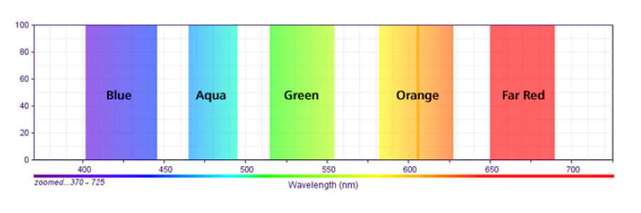
Typical fluorescence microscopy channels
Immunocytochemistry, immunohistochemistry and immunofluorescence—what are the differences?
Both immunocytochemistry (ICC) and immunohistochemistry (IHC) employ immune labeling of antigens for analysis and use immunofluorescence methods. Sometimes, immunocytochemistry and immunofluorescence are used interchangeably.
While ICC focuses on analysis at a cellular level, IHC allows examination of the whole tissue. Both techniques use immunofluorescence for detection. It is also possible to use enzyme-based detection (e.g., using 3,3’-diaminobenzidine (DAB)).
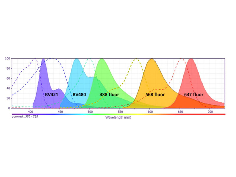
Robust and easy 5-color imaging
- Ability to quantitate fluorescence signals (as opposed to qualitative determination using enzyme-based methods)
- Ability to multiplex (fluorescent dyes with different emission spectra can be combined to detect multiple proteins)
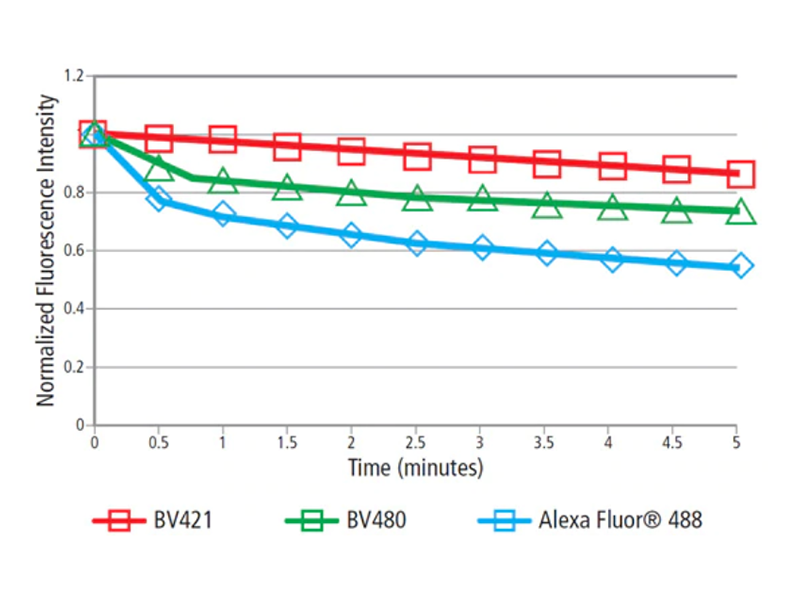
- Photostability of fluorescent dyes
Photostability of BV dyes
Right image: Continuous exposure to excitation light. Cells: MCF7 cells. Fixation: BD Cytofix™ Fixation Buffer (Cat. No. 554655). Antibodies: Purified Mouse Anti-Human CD324 (Cat No. 562869) with BD Horizon Brilliant Violet™ 480 (BV480) Goat Anti-Mouse (Cat. No.564877), BD Horizon Brilliant Violet™ 421 (BV421) Goat-Anti-Mouse (Cat. No. 563846), or Alexa Fluor™ 488 Goat Anti-Mouse (Thermo Fisher Scientific). Other reagents: ProLong™ Gold Antifade Mountant (Thermo Fisher Scientific). Instrument: ImageXpress™ Micro XLS System (Molecular Devices). 20X objective.
BD Biosciences immunofluorescence imaging product portfolio
BD Biosciences offers high-quality tools for fluorescence imaging, which include a comprehensive research portfolio of monoclonal antibodies, fluorescent dyes, second step reagents, and buffers. We offer protocols for immunofluorescence and for preparation and staining of tissue sections.
Immunofluorescence imaging protocols and resources
We also offer protocols to help support your sample preparation and immunofluorescence analysis.
BD Biosciences’ fluorescent dyes for imaging applications build on our experience in fluorescent imaging and use the polymer dye technology acquired from Sirigen Ltd. The exceptional brightness of our fluorophores enables better sensitivity. The unique spectra allow for easier multiplexing.
BD Horizon Brilliant™ Fluorophores
Learn more about BD Horizon Brilliant Violet™ Imaging Reagents
-
Recommended filter configuration when using epifluorescence microscopy and DAPI staining
Channel Dyes Excitation (nm) DC (nm) Emission (nm) Optimized Filter Sets Blue DAPI 377/50 409 430/24 or 440/40 Chroma or Semrock Aqua BD Horizon Brilliant
Violet™ 480 (BV480)445/20 or 438/24 458 483/32 Chroma or Semrock Green FITC, Alexa Fluor™ 488,
eGFP, zsGreen490/30 or 485/20 506 or 495 537/26 or 525/50 Chroma or Semrock Orange Alexa Fluor™ 555,
Alexa Fluor™ 568Any optimized filter set Any optimized filter set Any optimized filter set Any vendor Red Alexa Fluor™ 594 Any optimized filter set Any optimized filter set Any optimized filter set Any vendor Far Red Alexa Fluor™ 647 Any optimized filter set Any optimized filter set Any optimized filter set Any vendor Recommended filter configuration to adopt BD Horizon Brilliant Violet™ 480 (BV480) for improved immunofluorescence assays.
-
Recommended filter configuration when using epifluorescence and a nuclear counterstain other than DAPI
Configuration for one brighter dye: BD Horizon Brilliant Violet™ 421 (BV421) for improved immunofluorescence assays.
Channel Dyes Excitation (nm) DC (nm) Emission (nm) Optimized Filter Sets Blue BD Horizon Brilliant
Violet™ 421 (BV421)392/23 or 377/50 409 430/24 or 440/40 Chroma or Semrock Aqua BD Horizon Brilliant
Violet™ 480 (BV480)438/24 or 445/20 458 483/32 Chroma or Semrock Green FITC, Alexa Fluor™ 488,
eGFP, zsGreen490/30 or 485/20 506 or 495 537/26 or 525/50 Chroma or Semrock Orange Alexa Fluor™ 555,
Alexa Fluor™ 568Any optimized filter set Any optimized filter set Any optimized filter set Any vendor Red Alexa Fluor™ 594 Any optimized filter set Any optimized filter set Any optimized filter set Any vendor Far Red Alexa Fluor™ 647,
DRAQ5™ (DNA)Any optimized filter set Any optimized filter set Any optimized filter set Any vendor Recommended filter configuration to adopt BD Horizon Brilliant Violet™ 421 (BV421) for improved immunofluorescence assays.
Configuration for two brighter dyes: BD Horizon Brilliant Violet™ 421 (BV421) and BV480 for
improved immunofluorescence assays.Channel Dyes Excitation (nm) DC (nm) Emission (nm) Optimized Filter Sets Blue BD Horizon Brilliant
Violet™ 421 (BV421)392/23 or 377/50 409 430/24 or 440/40 Chroma or Semrock Aqua BD Horizon Brilliant
Violet™ 480 (BV480)438/24 or 445/20 458 483/32 Chroma or Semrock Green FITC, Alexa Fluor™ 488,
eGFP, zsGreen490/30 or 485/20 506 or 495 537/26 or 525/50 Chroma or Semrock Orange Alexa Fluor™ 555,
Alexa Fluor™ 568Any optimized filter set Any optimized filter set Any optimized filter set Any vendor Red Alexa Fluor™ 594 Any optimized filter set Any optimized filter set Any optimized filter set Any vendor Far Red Alexa Fluor™ 647,
DRAQ5™ (DNA)Any optimized filter set Any optimized filter set Any optimized filter set Any vendor -
Recommended filter configuration when using laser confocal microscopy
Up to 5-Color Configuration Laser Line Dyes DAPI Essential 405 nm DAPI 440 nm or 458 nm BD Horizon Brilliant Violet™ 480 (BV480) 488 nm Alexa Fluor™ 488 or GFP 561 nm Alexa Fluor™ 568 or RFP 633 nm Alexa Fluor™ 647 Alternate DNA Stain 405 nm BD Horizon Brilliant Violet™ 421 (BV421) and
BD Horizon Brilliant Violet™ 480 (BV480)440 nm or 458 nm Laser line not essential 488 nm Alexa Fluor™ 488 or GFP 561 nm Alexa Fluor™ 568 or RFP 633 nm DRAQ5™ or Alexa Fluor™ 647
Sample data generated using BD Biosciences antibodies and reagents
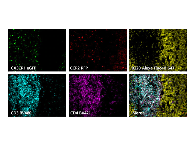
Spleen cryosections (5 µm) from mice expressing CX3CR1-green fluorescent protein (pseudo-colored green) and CCR2-red fluorescent protein (pseudo-colored red) were fixed with BD Cytofix™ Fixation Buffer (Cat. No. 554655), blocked with 5% goat serum and 1% BSA diluted in 1X PBS, and stained with BD Horizon Brilliant Violet™ 421 (BV421) Rat Anti-Mouse CD4 (Cat. No. 562891) (pseudo-colored magenta), Alexa Fluor™ 647 Rat Anti-Mouse CD45R/B220 (Cat No. 557683) (pseudo-colored yellow), and BD Horizon Brilliant Violet™ 480 (BV480) Rat Anti-Mouse CD3 Molecular Complex (pseudo-colored cyan). Slides were mounted with ProLong™ Gold Antifade Mountant (Thermo Fisher Scientific) and the image was captured on a BD Pathway 435 Cell Analyzer (epifluorescence microscope) and merged using BD Attovision Software. 20X objective. Carried out in collaboration with the Catherine Hedrick lab at La Jolla Institute of Allergy and Immunology.
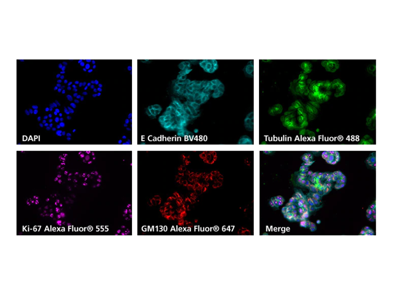
MCF-7 cells (ATCC, HTB-22) were fixed with BD Cytofix™ Fixation Buffer (Cat. No. 554655), permeabilized with 0.1% Triton™ X-100 and blocked with 5% goat serum, 1% BSA, 0.5% Triton™ X-100 in 1X PBS. Cells were incubated with Purified Mouse Anti-E-Cadherin (Cat. No. 610182) and the second-step reagent was BD Horizon Brilliant Violet™ 480 (BV480) Goat Anti-Mouse (Cat. No. 564877) (pseudo-colored aqua). Cells were then stained with directly conjugated antibodies: Alexa Fluor™ 488 Mouse Anti-β-Tubulin (Cat No. 558605) (pseudo-colored green), Alexa Fluor™ 555 Mouse Anti-Human Ki-67 (Cat. No. 558617) (pseudo-colored purple), Alexa Fluor™ 647 Mouse Anti-GM130 (Cat. No. 558712) (pseudo-colored red). DAPI (Cat. No. 564907) was used as a nuclear counterstain (pseudo-colored blue). Slides were mounted with ProLong™ Gold Antifade Mountant (Thermo Fisher Scientific) and the image was captured on a BD Pathway 435 Cell Analyzer and merged using BD Attovision Software (BD Biosciences). 20X objective.
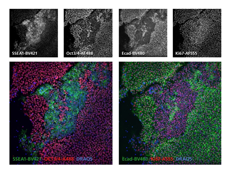
Cultured cells from the H9 human ES cell line were fixed with BD Cytofix™ Fixation Buffer (Cat. No. 554655), permeabilized with 0.1% Triton™ X-100 diluted in 1X PBS, blocked with 5% goat serum, 1% BSA, and 0.5% Triton™ X-100 diluted in 1X PBS, and stained with BD Horizon Brilliant Violet™ 421 (BV421) Mouse Anti-SSEA-1 (Cat. No. 562705), BD Horizon Brilliant Violet™ 480 (BV480) Mouse Anti-CD324(E-Cadherin) (Cat. No. 565646), Alexa Fluor™ 488 Mouse Anti-Oct3/4 (Cat. No. 561628), and Alexa Fluor™ 555 Mouse Anti-Human Ki-67 (Cat No. 558617). DRAQ5™ (Cat. No. 564902) was used as a nuclear counterstain. Slides were mounted with ProLong™ Gold Antifade Mountant (Thermo Fisher Scientific) and the images were captured on a BD Pathway 435 Cell Analyzer (epifluorescence microscope) and merged using BD Attovision Software. 20X objective.
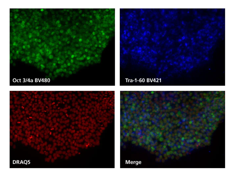
Cells: H9 human embryonic stem cells (WiCell, Madison WI). Fix: BD Cytofix™ Fixation Buffer (Cat. No. 554655); Perm: BD Phosflow™ Perm Buffer III (Cat. No. 558050), Block/Stain: 5% goat serum, 1% BSA, 0.5 % Triton™ X-100 in 1X PBS. Antibodies: Purified Mouse Anti-Oct3/4a (Cat. No. 561555) with BD Horizon Brilliant Violet™ 480 (BV480) Goat Anti-Mouse (Cat. No. 564877), BD Horizon Brilliant Violet™ 421 (BV421) Mouse Anti-Human TRA-1-60 Antigen (Cat. No. 562711). Other reagents: DRAQ5™ (BioStatus), ProLong™ Gold Antifade Mountant (Thermo Fisher Scientific). Instrument: BD Pathway 435 Cell Analyzer and images merged using BD Attovision Software (BD Biosciences). 20X objective.
Applications using BD Biosciences immunofluorescence reagents
See how BD Biosciences immunofluorescence reagents were used in various imaging applications.
For Research Use Only. Not for use in diagnostic or therapeutic procedures.
Alexa Fluor and ProLong are trademarks of Thermo Fisher Scientific.
DRAQ5 is a trademark of BioStatus Limited. ImageXpress is a trademark of Molecular Devices.
Triton is a trademark of Dow.