-
Reagents
- Flow Cytometry Reagents
-
Western Blotting and Molecular Reagents
- Immunoassay Reagents
-
Single-Cell Multiomics Reagents
- BD® OMICS-Guard Sample Preservation Buffer
- BD® AbSeq Assay
- BD® Single-Cell Multiplexing Kit
- BD Rhapsody™ ATAC-Seq Assays
- BD Rhapsody™ Whole Transcriptome Analysis (WTA) Amplification Kit
- BD Rhapsody™ TCR/BCR Next Multiomic Assays
- BD Rhapsody™ Targeted mRNA Kits
- BD Rhapsody™ Accessory Kits
- BD® OMICS-One Protein Panels
-
Functional Assays
-
Microscopy and Imaging Reagents
-
Cell Preparation and Separation Reagents
-
- BD® OMICS-Guard Sample Preservation Buffer
- BD® AbSeq Assay
- BD® Single-Cell Multiplexing Kit
- BD Rhapsody™ ATAC-Seq Assays
- BD Rhapsody™ Whole Transcriptome Analysis (WTA) Amplification Kit
- BD Rhapsody™ TCR/BCR Next Multiomic Assays
- BD Rhapsody™ Targeted mRNA Kits
- BD Rhapsody™ Accessory Kits
- BD® OMICS-One Protein Panels
- Ireland (English)
-
Change country/language
Old Browser
This page has been recently translated and is available in French now.
Looks like you're visiting us from United States.
Would you like to stay on the current country site or be switched to your country?
BD Transduction Laboratories™ Purified Mouse Anti-JAK1
Clone 73/JAK1 (RUO)


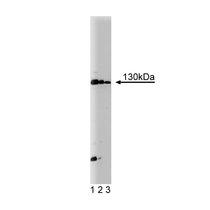
Western blot analysis of JAK1 on Jurkat cell lysate. Lane 1: 1:250, lane 2: 1:500, lane 3: 1:1000 dilution of JAK1.

Immunofluorescent staining of human endothelial cells.


BD Transduction Laboratories™ Purified Mouse Anti-JAK1

BD Transduction Laboratories™ Purified Mouse Anti-JAK1

Regulatory Status Legend
Any use of products other than the permitted use without the express written authorization of Becton, Dickinson and Company is strictly prohibited.
Preparation And Storage
Product Notices
- Since applications vary, each investigator should titrate the reagent to obtain optimal results.
- Please refer to www.bdbiosciences.com/us/s/resources for technical protocols.
- Source of all serum proteins is from USDA inspected abattoirs located in the United States.
- Caution: Sodium azide yields highly toxic hydrazoic acid under acidic conditions. Dilute azide compounds in running water before discarding to avoid accumulation of potentially explosive deposits in plumbing.
- For fluorochrome spectra and suitable instrument settings, please refer to our Multicolor Flow Cytometry web page at www.bdbiosciences.com/colors.
The JAK family of receptor-associated protein kinases is directly involved in interferon (IFN) response pathways. The JAK family contains at least three members: JAK1, JAK2, and Tyk2. Each protein is approximately 130 kDa and contains a C-terminal tyrosine kinase domain, an adjacent kinase or kinase-related domain, and five other domains that are highly conserved among family members. In several human and murine cell lines, JAK1 is rapidly tyrosine phosphorylated in response to IFN-α and IFN-γ. JAK1 is required for the phosphorylation of the transcription factor Stat1 (p91), in response to IFNs-α or IFN-γ. JAK1 is also necessary for the efficient phophorylation of Stat2 (p113) in response to IFN-α and for the phosphorylation of Tyk2 or JAK2 in response to IFNs-α or IFN-γ, respectively.
Development References (5)
-
Blesofsky WA, Mowen K, Arduini RM. Regulation of STAT protein synthesis by c-Cbl. Oncogene. 2001; 20(50):7326-7333. (Clone-specific: Immunoprecipitation, Western blot). View Reference
-
Kawazoe Y, Naka T, Fujimoto M. Signal transducer and activator of transcription (STAT)-induced STAT inhibitor 1 (SSI-1)/suppressor of cytokine signaling 1 (SOCS1) inhibits insulin signal transduction pathway through modulating insulin receptor substrate 1 (IRS-1) phosphorylation. J Exp Med. 2001; 193(2):263-269. (Clone-specific: Western blot). View Reference
-
Kopantzev Y, Heller M, Swaminathan N, Rudikoff S. IL-6 mediated activation of STAT3 bypasses Janus kinases in terminally differentiated B lineage cells. Oncogene. 2002; 21(44):6791-6800. (Clone-specific: Immunoprecipitation, Western blot). View Reference
-
Muller M, Briscoe J, Laxton C. The protein tyrosine kinase JAK1 complements defects in interferon-alpha/beta and -gamma signal transduction. Nature. 1993; 366(6451):129-135. (Biology). View Reference
-
Nicholson SE, Oates AC, Harpur AG, Ziemiecki A, Wilks AF, Layton JE. Tyrosine kinase JAK1 is associated with the granulocyte-colony-stimulating factor receptor and both become tyrosine-phosphorylated after receptor activation. Proc Natl Acad Sci U S A. 1994; 91(8):2985-2988. (Biology). View Reference
Please refer to Support Documents for Quality Certificates
Global - Refer to manufacturer's instructions for use and related User Manuals and Technical data sheets before using this products as described
Comparisons, where applicable, are made against older BD Technology, manual methods or are general performance claims. Comparisons are not made against non-BD technologies, unless otherwise noted.
For Research Use Only. Not for use in diagnostic or therapeutic procedures.