-
Reagents
- Flow Cytometry Reagents
-
Western Blotting and Molecular Reagents
- Immunoassay Reagents
-
Single-Cell Multiomics Reagents
- BD® OMICS-Guard Sample Preservation Buffer
- BD® AbSeq Assay
- BD® Single-Cell Multiplexing Kit
- BD Rhapsody™ ATAC-Seq Assays
- BD Rhapsody™ Whole Transcriptome Analysis (WTA) Amplification Kit
- BD Rhapsody™ TCR/BCR Next Multiomic Assays
- BD Rhapsody™ Targeted mRNA Kits
- BD Rhapsody™ Accessory Kits
- BD® OMICS-One Protein Panels
-
Functional Assays
-
Microscopy and Imaging Reagents
-
Cell Preparation and Separation Reagents
-
- BD® OMICS-Guard Sample Preservation Buffer
- BD® AbSeq Assay
- BD® Single-Cell Multiplexing Kit
- BD Rhapsody™ ATAC-Seq Assays
- BD Rhapsody™ Whole Transcriptome Analysis (WTA) Amplification Kit
- BD Rhapsody™ TCR/BCR Next Multiomic Assays
- BD Rhapsody™ Targeted mRNA Kits
- BD Rhapsody™ Accessory Kits
- BD® OMICS-One Protein Panels
- Ireland (English)
-
Change country/language
Old Browser
This page has been recently translated and is available in French now.
Looks like you're visiting us from United States.
Would you like to stay on the current country site or be switched to your country?
BD Transduction Laboratories™ Purified Mouse Anti-Human PKCθ
Clone 27/PKCθ (RUO)


Western blot analysis of PKCθ on a Jurkat lysate. Lane 1: 1:250, lane 2: 1:500, lane 3: 1:1000 dilution of the anti- human PKCθ antibody.

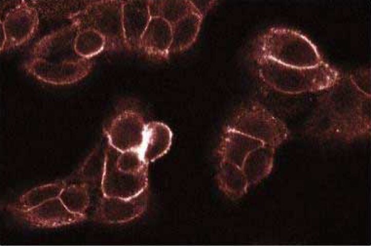
Immunofluorescence staining of A431 cells.


BD Transduction Laboratories™ Purified Mouse Anti-Human PKCθ

BD Transduction Laboratories™ Purified Mouse Anti-Human PKCθ

Regulatory Status Legend
Any use of products other than the permitted use without the express written authorization of Becton, Dickinson and Company is strictly prohibited.
Preparation And Storage
Product Notices
- Since applications vary, each investigator should titrate the reagent to obtain optimal results.
- Please refer to www.bdbiosciences.com/us/s/resources for technical protocols.
- Caution: Sodium azide yields highly toxic hydrazoic acid under acidic conditions. Dilute azide compounds in running water before discarding to avoid accumulation of potentially explosive deposits in plumbing.
- Source of all serum proteins is from USDA inspected abattoirs located in the United States.
Data Sheets
Companion Products

.png?imwidth=320)

The Protein Kinase C (PKC) family of homologous serine/threonine protein kinases is involved in a number of processes, such as growth, differentiation, and cytokine secretion. At least eleven isozymes have been described. These proteins are products of multiple genes and alternative splicing. Conventional PKC (cPKC) subfamily members (α, β, and γ isoforms) consists of a single polypeptide chain containing four conserved regions (C) and five variable regions (V). The N-terminal half containing C1, C2, V1, and V2 constitutes the regulatory domain and interacts with the PKC activators Ca2+, phospholipid, diacylglycerol, or phorbol ester. However, the the C2-like domains of novel PKC (nPKC) subfamily members (δ, ε, η, and θ isoforms) are Ca2+-independent. The atypical PKC (aPKC) subfamily members (ζ, ι, and λ isoforms) lack the C2 domain and are unique in that their activity is independent of diacylglycerols and phorbol esters. They also lack one repeat of the cysteine-rich sequences that are conserved in cPKC and nPKC members. The C-terminal region of PKC contains the catalytic domain. The PKC pathway represents a major signal transduction system that is activated following ligand-stimulation of transmembrane receptors by hormones, neurotransmitters, and growth factors. PKCθ transcripts are expressed in most tissues with the highest levels being found in hematopoietic tissues and cell lines, including T cells and thymocytes. PKCθ mRNA is readily detectable in skeletal muscle, lung and brain. However, PKCθ expression is not detected in several human carcinoma cell lines. Abundant expression of this PKC isozyme in hematopoietic cells suggest that it may have a role in growth and differentiation processes of these cells.
The 27/PKCθ monoclonal antibody recognizes the C2-like domain of human PKCθ, regardless of phosphorylation status.
This antibody is routinely tested by western blot analysis. Other applications were tested at BD Biosciences Pharmingen during antibody development only or reported in the literature.
Development References (5)
-
Dienz O, Hehner SP, Droge W, Schmitz ML. Synergistic activation of NF-kappa B by functional cooperation between vav and PKCtheta in T lymphocytes. J Biol Chem. 2000; 275(32):24547-24551. (Biology: Immunoprecipitation, Western blot). View Reference
-
Nishizuka Y. The molecular heterogeneity of protein kinase C and its implications for cellular regulation. Nature. 1988; 334(6184):661-665. (Biology). View Reference
-
Soderling TR. Protein kinases. Regulation by autoinhibitory domains. J Biol Chem. 1990; 265(4):1823-1826. (Biology). View Reference
-
Villalba M, Bi K, Rodriguez F, Tanaka Y, Schoenberger S, Altman A. Vav1/Rac-dependent actin cytoskeleton reorganization is required for lipid raft clustering in T cells. J Cell Biol. 2001; 155(3):331-338. (Biology: Immunofluorescence). View Reference
-
Villalba M, Coudronniere N, Deckert M, Teixeiro E, Mas P, Altman A. A novel functional interaction between Vav and PKCtheta is required for TCR-induced T cell activation. Immunity. 2000; 12(2):151-160. (Biology: Immunoprecipitation, In vitro kinase assay, Western blot). View Reference
Please refer to Support Documents for Quality Certificates
Global - Refer to manufacturer's instructions for use and related User Manuals and Technical data sheets before using this products as described
Comparisons, where applicable, are made against older BD Technology, manual methods or are general performance claims. Comparisons are not made against non-BD technologies, unless otherwise noted.
For Research Use Only. Not for use in diagnostic or therapeutic procedures.