-
Reagents
- Flow Cytometry Reagents
-
Western Blotting and Molecular Reagents
- Immunoassay Reagents
-
Single-Cell Multiomics Reagents
- BD® OMICS-Guard Sample Preservation Buffer
- BD® AbSeq Assay
- BD® Single-Cell Multiplexing Kit
- BD Rhapsody™ ATAC-Seq Assays
- BD Rhapsody™ Whole Transcriptome Analysis (WTA) Amplification Kit
- BD Rhapsody™ TCR/BCR Next Multiomic Assays
- BD Rhapsody™ Targeted mRNA Kits
- BD Rhapsody™ Accessory Kits
- BD® OMICS-One Protein Panels
-
Functional Assays
-
Microscopy and Imaging Reagents
-
Cell Preparation and Separation Reagents
-
- BD® OMICS-Guard Sample Preservation Buffer
- BD® AbSeq Assay
- BD® Single-Cell Multiplexing Kit
- BD Rhapsody™ ATAC-Seq Assays
- BD Rhapsody™ Whole Transcriptome Analysis (WTA) Amplification Kit
- BD Rhapsody™ TCR/BCR Next Multiomic Assays
- BD Rhapsody™ Targeted mRNA Kits
- BD Rhapsody™ Accessory Kits
- BD® OMICS-One Protein Panels
- Ireland (English)
-
Change country/language
Old Browser
This page has been recently translated and is available in French now.
Looks like you're visiting us from United States.
Would you like to stay on the current country site or be switched to your country?
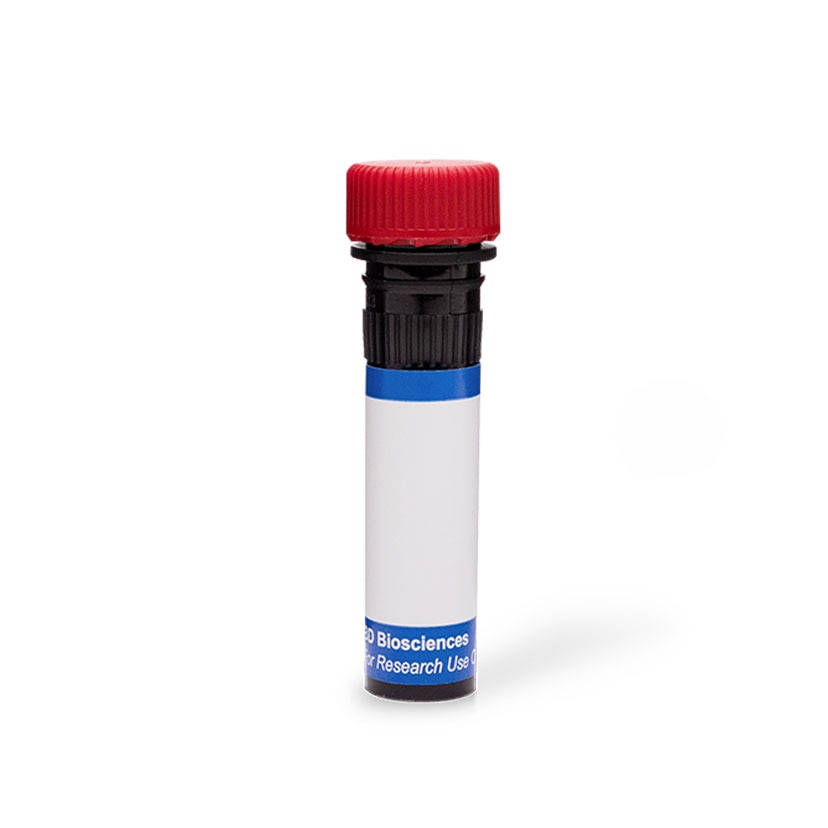
BD Pharmingen™ PE Rat Anti-Mouse CD115 (CSF-1R)
Clone AFS98 (also known as AFS98) (RUO)

Two-color flow cytometric analysis of CD115 (CSF-1R) expression on mouse bone marrow cells. C57BL/6 mouse bone marrow cells were harvested and erythrocytes were lysed with BD Pharm Lyse™ Lysing Buffer (Cat. No. 555899). The cells were then washed and stained with Alexa Fluor® 647 Rat Anti-Mouse CD11b antibody (Cat. No. 557686) and either PE Rat IgG2a, κ Isotype Control (Cat. No. 553930; Left Plot) or PE Rat Anti-Mouse CD115 (CSF-1R) antibody (Cat. No. 566839; Right Plot) at 0.25 µg/test. DAPI (4',6-Diamidino-2-Phenylindole, Dihydrochloride) Solution (Cat. No. 564907) was added to cells right before analysis. Two-color flow cytometric contour plots showing the correlated expression of CD115 (CSF-1R) [or Ig Isotype control staining] versus CD11b were derived from gated events with the forward and side-light scattering characteristics of viable (DAPI-negative) bone marrow cells. Flow cytometry and data analysis were performed using a BD LSRFortessa™ X-20 Cell Analyzer System and FlowJo™ software. Data shown in this Technical Data Sheet are not lot specific.

Two-color flow cytometric analysis of CD115 (CSF-1R) expression on mouse bone marrow cells. C57BL/6 mouse bone marrow cells were harvested and erythrocytes were lysed with BD Pharm Lyse™ Lysing Buffer (Cat. No. 555899). The cells were then washed and stained with Alexa Fluor® 647 Rat Anti-Mouse CD11b antibody (Cat. No. 557686) and either PE Rat IgG2a, κ Isotype Control (Cat. No. 553930; Left Plot) or PE Rat Anti-Mouse CD115 (CSF-1R) antibody (Cat. No. 566839; Right Plot) at 0.25 µg/test. DAPI (4',6-Diamidino-2-Phenylindole, Dihydrochloride) Solution (Cat. No. 564907) was added to cells right before analysis. Two-color flow cytometric contour plots showing the correlated expression of CD115 (CSF-1R) [or Ig Isotype control staining] versus CD11b were derived from gated events with the forward and side-light scattering characteristics of viable (DAPI-negative) bone marrow cells. Flow cytometry and data analysis were performed using a BD LSRFortessa™ X-20 Cell Analyzer System and FlowJo™ software. Data shown in this Technical Data Sheet are not lot specific.


Two-color flow cytometric analysis of CD115 (CSF-1R) expression on mouse bone marrow cells. C57BL/6 mouse bone marrow cells were harvested and erythrocytes were lysed with BD Pharm Lyse™ Lysing Buffer (Cat. No. 555899). The cells were then washed and stained with Alexa Fluor® 647 Rat Anti-Mouse CD11b antibody (Cat. No. 557686) and either PE Rat IgG2a, κ Isotype Control (Cat. No. 553930; Left Plot) or PE Rat Anti-Mouse CD115 (CSF-1R) antibody (Cat. No. 566839; Right Plot) at 0.25 µg/test. DAPI (4',6-Diamidino-2-Phenylindole, Dihydrochloride) Solution (Cat. No. 564907) was added to cells right before analysis. Two-color flow cytometric contour plots showing the correlated expression of CD115 (CSF-1R) [or Ig Isotype control staining] versus CD11b were derived from gated events with the forward and side-light scattering characteristics of viable (DAPI-negative) bone marrow cells. Flow cytometry and data analysis were performed using a BD LSRFortessa™ X-20 Cell Analyzer System and FlowJo™ software. Data shown in this Technical Data Sheet are not lot specific.
Two-color flow cytometric analysis of CD115 (CSF-1R) expression on mouse bone marrow cells. C57BL/6 mouse bone marrow cells were harvested and erythrocytes were lysed with BD Pharm Lyse™ Lysing Buffer (Cat. No. 555899). The cells were then washed and stained with Alexa Fluor® 647 Rat Anti-Mouse CD11b antibody (Cat. No. 557686) and either PE Rat IgG2a, κ Isotype Control (Cat. No. 553930; Left Plot) or PE Rat Anti-Mouse CD115 (CSF-1R) antibody (Cat. No. 566839; Right Plot) at 0.25 µg/test. DAPI (4',6-Diamidino-2-Phenylindole, Dihydrochloride) Solution (Cat. No. 564907) was added to cells right before analysis. Two-color flow cytometric contour plots showing the correlated expression of CD115 (CSF-1R) [or Ig Isotype control staining] versus CD11b were derived from gated events with the forward and side-light scattering characteristics of viable (DAPI-negative) bone marrow cells. Flow cytometry and data analysis were performed using a BD LSRFortessa™ X-20 Cell Analyzer System and FlowJo™ software. Data shown in this Technical Data Sheet are not lot specific.

Two-color flow cytometric analysis of CD115 (CSF-1R) expression on mouse bone marrow cells. C57BL/6 mouse bone marrow cells were harvested and erythrocytes were lysed with BD Pharm Lyse™ Lysing Buffer (Cat. No. 555899). The cells were then washed and stained with Alexa Fluor® 647 Rat Anti-Mouse CD11b antibody (Cat. No. 557686) and either PE Rat IgG2a, κ Isotype Control (Cat. No. 553930; Left Plot) or PE Rat Anti-Mouse CD115 (CSF-1R) antibody (Cat. No. 566839; Right Plot) at 0.25 µg/test. DAPI (4',6-Diamidino-2-Phenylindole, Dihydrochloride) Solution (Cat. No. 564907) was added to cells right before analysis. Two-color flow cytometric contour plots showing the correlated expression of CD115 (CSF-1R) [or Ig Isotype control staining] versus CD11b were derived from gated events with the forward and side-light scattering characteristics of viable (DAPI-negative) bone marrow cells. Flow cytometry and data analysis were performed using a BD LSRFortessa™ X-20 Cell Analyzer System and FlowJo™ software. Data shown in this Technical Data Sheet are not lot specific.

Two-color flow cytometric analysis of CD115 (CSF-1R) expression on mouse bone marrow cells. C57BL/6 mouse bone marrow cells were harvested and erythrocytes were lysed with BD Pharm Lyse™ Lysing Buffer (Cat. No. 555899). The cells were then washed and stained with Alexa Fluor® 647 Rat Anti-Mouse CD11b antibody (Cat. No. 557686) and either PE Rat IgG2a, κ Isotype Control (Cat. No. 553930; Left Plot) or PE Rat Anti-Mouse CD115 (CSF-1R) antibody (Cat. No. 566839; Right Plot) at 0.25 µg/test. DAPI (4',6-Diamidino-2-Phenylindole, Dihydrochloride) Solution (Cat. No. 564907) was added to cells right before analysis. Two-color flow cytometric contour plots showing the correlated expression of CD115 (CSF-1R) [or Ig Isotype control staining] versus CD11b were derived from gated events with the forward and side-light scattering characteristics of viable (DAPI-negative) bone marrow cells. Flow cytometry and data analysis were performed using a BD LSRFortessa™ X-20 Cell Analyzer System and FlowJo™ software. Data shown in this Technical Data Sheet are not lot specific.


Regulatory Status Legend
Any use of products other than the permitted use without the express written authorization of Becton, Dickinson and Company is strictly prohibited.
Preparation And Storage
Product Notices
- Since applications vary, each investigator should titrate the reagent to obtain optimal results.
- An isotype control should be used at the same concentration as the antibody of interest.
- Caution: Sodium azide yields highly toxic hydrazoic acid under acidic conditions. Dilute azide compounds in running water before discarding to avoid accumulation of potentially explosive deposits in plumbing.
- For fluorochrome spectra and suitable instrument settings, please refer to our Multicolor Flow Cytometry web page at www.bdbiosciences.com/colors.
- Please refer to http://regdocs.bd.com to access safety data sheets (SDS).
- Please refer to www.bdbiosciences.com/us/s/resources for technical protocols.
Data Sheets
Companion Products


Recently Viewed
The AFS98 monoclonal antibody specifically recognizes CD115 which is also known as Colony stimulating factor 1 Receptor (CSF-1R), Macrophage colony-stimulating factor 1 receptor (M-CSFR), or c-fms. This type I transmembrane glycoprotein is a receptor tyrosine kinase (RTK) that is encoded by Csf1r which belongs to the Ig superfamily. CD115 (CSF-1R) is comprised of an extracellular ligand-binding domain that is followed by a single transmembrane segment and a split intracellular tyrosine kinase domain. This receptor is expressed on monocytes, macrophages, dendritic cells, osteoclasts, and their precursors. Colony-stimulating factor 1 (CSF-1), also known as Macrophage colony-stimulating factor (M-CSF), binds to and signals through CD115 (CSF-1R) homodimers which undergo tyrosine autophosphorylation. The activated receptors transduce intracellular signals resulting in cytoskeletal reorganization and gene expression involved in the proliferation, differentiation, and survival of CSF-1-responding cells. Through CD115 (CSF-1R), CSF-1 regulates the release of proinflammatory cytokines and other mediators from macrophages and plays a role in the bone resorption activity of osteoclasts. Interleukin-34 (IL-34) is another ligand for CD115 (CSF-1R) that can induce similar, as well as, some different biological responses by target cells that express this receptor.

Development References (5)
-
Fend L, Accart N, Kintz J et al. Therapeutic effects of anti-CD115 monoclonal antibody in mouse cancer models through dual inhibition of tumor-associated macrophages and osteoclasts. PLoS ONE. 2013; 8(9):e73310. (Clone-specific: Blocking). View Reference
-
Jose MD, Le Meur Y, Atkins RC, Chadban SJ. Blockade of macrophage colony-stimulating factor reduces macrophage proliferation and accumulation in renal allograft rejection.. Am J Transplant. 2003; 3(3):294-300. (Clone-specific: Blocking, Immunohistochemistry, Inhibition). View Reference
-
Murayama T, Yokode M, Kataoka H, et al. Intraperitoneal administration of anti-c-fms monoclonal antibody prevents initial events of atherogenesis but does not reduce the size of advanced lesions in apolipoprotein E-deficient mice.. Circulation. 1999; 99(13):1740-6. (Clone-specific: Flow cytometry, In vivo exacerbation). View Reference
-
Rothwell VM, Rohrschneider LR. Murine c-fms cDNA: cloning, sequence analysis and retroviral expression. Oncogene Res. 1987; 1(4):311-324. (Biology). View Reference
-
Sudo T, Nishikawa S, Ogawa M, et al. Functional hierarchy of c-kit and c-fms in intramarrow production of CFU-M.. Oncogene. 1995; 11(12):2469-76. (Immunogen: Blocking, Flow cytometry, Fluorescence activated cell sorting, Functional assay, Immunoprecipitation, Inhibition, In vivo exacerbation, Radioimmunoassay). View Reference
Please refer to Support Documents for Quality Certificates
Global - Refer to manufacturer's instructions for use and related User Manuals and Technical data sheets before using this products as described
Comparisons, where applicable, are made against older BD Technology, manual methods or are general performance claims. Comparisons are not made against non-BD technologies, unless otherwise noted.
For Research Use Only. Not for use in diagnostic or therapeutic procedures.
