





.png)


Preparation And Storage


Product Notices
- Since applications vary, each investigator should titrate the reagent to obtain optimal results.
- An isotype control should be used at the same concentration as the antibody of interest.
- Caution: Sodium azide yields highly toxic hydrazoic acid under acidic conditions. Dilute azide compounds in running water before discarding to avoid accumulation of potentially explosive deposits in plumbing.
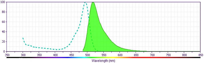
- For fluorochrome spectra and suitable instrument settings, please refer to our Multicolor Flow Cytometry web page at www.bdbiosciences.com/colors.
- Please refer to www.bdbiosciences.com/us/s/resources for technical protocols.
Companion Products


.png?imwidth=320)
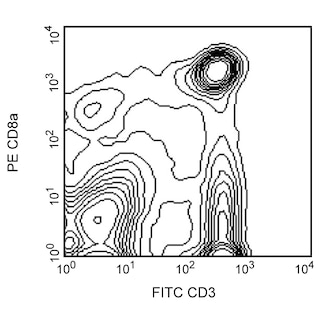
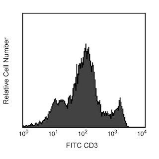
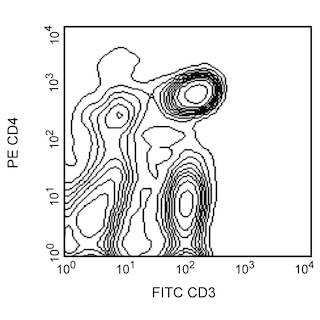
The G4.18 monoclonal antibody specifically recognizes the T-cell receptor-associated CD3 cell-surface antigen found on thymocytes, peripheral T lymphocytes, and dendritic epidermal T cells. It has been reported that CD3 expression is down-regulated within 24 hours in concanavalin A-stimulated rat T cells, and soluble mAb inhibits the allogeneic mixed-lymphocyte proliferative response and cell-mediated cytotoxicity to allogeneic target cells. In vivo treatment with G4.18 mAb prevents cardiac and skin allograft rejection, resulting in donor-specific tolerance. Pre-incubation of splenocytes with the alternate anti-rat CD3 monoclonal antibody, 1F4, blocks staining with mAb G4.18.
Development References (5)
-
Morris DL, Komocsar WJ. Immunophenotyping analysis of peripheral blood, splenic, and thymic lymphocytes in male and female rats. J Pharmacol Toxicol Methods. 1997; 37(1):37-46. (Clone-specific). View Reference
-
Nelson DJ, McMenamin C, McWilliam AS, Brenan M, Holt PG. Development of the airway intraepithelial dendritic cell network in the rat from class II major histocompatibility (Ia)-negative precursors: differential regulation of Ia expression at different levels of the respiratory tract. J Exp Med. 1994; 179(1):203-212. (Clone-specific: Immunohistochemistry). View Reference
-
Nicolls MR, Aversa GG, Pearce NW, et al. Induction of long-term specific tolerance to allografts in rats by therapy with an anti-CD3-like monoclonal antibody.. Transplantation. 1993; 55(3):459-68. (Immunogen: (Co)-stimulation, Flow cytometry, Immunohistochemistry, Immunoprecipitation, Inhibition, Stimulation). View Reference
-
Strickland D, Kees UR, Holt PG. Regulation of T-cell activation in the lung: alveolar macrophages induce reversible T-cell anergy in vitro associated with inhibition of interleukin-2 receptor signal transduction. Immunology. 1996; 87(2):250-258. (Biology). View Reference
-
Upham JW, Strickland DH, Bilyk N, Robinson BW, Holt PG. Alveolar macrophages from humans and rodents selectively inhibit T-cell proliferation but permit T-cell activation and cytokine secretion. Immunology. 1995; 84(1):142-147. (Biology). View Reference
Please refer to Support Documents for Quality Certificates
Global - Refer to manufacturer's instructions for use and related User Manuals and Technical data sheets before using this products as described
Comparisons, where applicable, are made against older BD Technology, manual methods or are general performance claims. Comparisons are not made against non-BD technologies, unless otherwise noted.
For Research Use Only. Not for use in diagnostic or therapeutic procedures.
