Dear Customer, If you experience any text readability or display issues on our website, please update your web browser to the latest version
.
See More
Dear Customer, If you experience any text readability or display issues on our website, please update your web browser to the latest version
.
See MoreDear Customer, If you experience any text readability or display issues on our website, please update your web browser to the latest version
.
This page has been recently translated and is available in French now.
Looks like you're visiting us from {countryName}.
Would you like to stay on the current location site or be switched to your location?
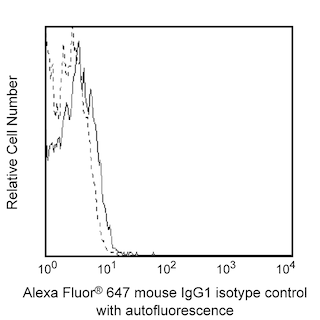
Flow cytometric analysis of fibronectin in human mesenchymal stem cells (MSC). MSC (Lonza), passage 6, were dissociated and fixed in BD Cytofix™ Fixation Buffer (Cat. No. 554655), and permeabilized with BD Phosflow™ Perm Buffer III (Cat. No. 558050). The cells were stained with either Alexa Fluor® 647 Mouse IgG1, κ isotype control (dashed line, Cat. No. 557714) or Alexa Fluor® 647 Anti-Fibronectin monoclonal antibody (solid line) at matched concentrations. Histograms were derived from gated events based on light scattering characteristics of MSC. Flow cytometry was performed on a BD LSRFortessa™ II flow cytometry system. BD Phosflow™ Perm/Wash Buffer I (Cat. No. 557885) is also suitable for permeabilization.
Immunofluorescent analysis of fibronectin in human mesenchymal stem cells (MSC). MSC (Lonza), passage 6, were fixed in BD Cytofix™ Fixation Buffer (Cat. No. 554655), permeabilized with 0.1% Triton™ X-100, and stained with Alexa Fluor® 647 Mouse Anti-Fibronectin monoclonal antibody (pseudo-colored red) at 1.2 µg/ml. Cell nuclei were stained with DAPI (pseudo-colored blue). The images were captured on a BD Pathway™ 435 Cell Analyzer and merged using BD Attovision™Software. BD Phosflow™ Perm Buffer III (Cat. No. 558050) is also suitable for permeabilization.
Flow cytometric analysis of fibronectin in human mesenchymal stem cells (MSC). MSC (Lonza), passage 6, were dissociated and fixed in BD Cytofix™ Fixation Buffer (Cat. No. 554655), and permeabilized with BD Phosflow™ Perm Buffer III (Cat. No. 558050). The cells were stained with either Alexa Fluor® 647 Mouse IgG1, κ isotype control (dashed line, Cat. No. 557714) or Alexa Fluor® 647 Anti-Fibronectin monoclonal antibody (solid line) at matched concentrations. Histograms were derived from gated events based on light scattering characteristics of MSC. Flow cytometry was performed on a BD LSRFortessa™ II flow cytometry system. BD Phosflow™ Perm/Wash Buffer I (Cat. No. 557885) is also suitable for permeabilization.
Immunofluorescent analysis of fibronectin in human mesenchymal stem cells (MSC). MSC (Lonza), passage 6, were fixed in BD Cytofix™ Fixation Buffer (Cat. No. 554655), permeabilized with 0.1% Triton™ X-100, and stained with Alexa Fluor® 647 Mouse Anti-Fibronectin monoclonal antibody (pseudo-colored red) at 1.2 µg/ml. Cell nuclei were stained with DAPI (pseudo-colored blue). The images were captured on a BD Pathway™ 435 Cell Analyzer and merged using BD Attovision™Software. BD Phosflow™ Perm Buffer III (Cat. No. 558050) is also suitable for permeabilization.
Flow cytometric analysis of fibronectin in human mesenchymal stem cells (MSC). MSC (Lonza), passage 6, were dissociated and fixed in BD Cytofix™ Fixation Buffer (Cat. No. 554655), and permeabilized with BD Phosflow™ Perm Buffer III (Cat. No. 558050). The cells were stained with either Alexa Fluor® 647 Mouse IgG1, κ isotype control (dashed line, Cat. No. 557714) or Alexa Fluor® 647 Anti-Fibronectin monoclonal antibody (solid line) at matched concentrations. Histograms were derived from gated events based on light scattering characteristics of MSC. Flow cytometry was performed on a BD LSRFortessa™ II flow cytometry system. BD Phosflow™ Perm/Wash Buffer I (Cat. No. 557885) is also suitable for permeabilization.
Immunofluorescent analysis of fibronectin in human mesenchymal stem cells (MSC). MSC (Lonza), passage 6, were fixed in BD Cytofix™ Fixation Buffer (Cat. No. 554655), permeabilized with 0.1% Triton™ X-100, and stained with Alexa Fluor® 647 Mouse Anti-Fibronectin monoclonal antibody (pseudo-colored red) at 1.2 µg/ml. Cell nuclei were stained with DAPI (pseudo-colored blue). The images were captured on a BD Pathway™ 435 Cell Analyzer and merged using BD Attovision™Software. BD Phosflow™ Perm Buffer III (Cat. No. 558050) is also suitable for permeabilization.
Any use of products other than the permitted use without the express written authorization of Becton, Dickinson and Company is strictly prohibited.
The 240-kDa dimeric fibronectin protein exists in two forms: a soluble protomer in body fluids and an insoluble multimer in the extracellular matrix. The latter is the primary functional form and creates a substrate for cell migration, a role which makes fibronectin vital to embryogenesis and wound response. Fibronectin mediates cytoskeletal organization, cell attachment, and cellular signaling through interactions with cellular receptors. Although various isoforms of fibronectin are derived by alternative splicing, they share a common N-terminus which is a critical region for cell surface binding in an initial step of multimer assembly. Further polymerization steps are regulated by fibronectin/integrin interactions and result in generation of the complex fibrils that constitute the fibronectin matrix.
Please refer to Support Documents for Quality Certificates
Global - Refer to manufacturer's instructions for use and related User Manuals and Technical data sheets before using this products as described
Comparisons, where applicable, are made against older BD Technology, manual methods or are general performance claims. Comparisons are not made against non-BD technologies, unless otherwise noted.
Please refer to Support Documents for Quality Certificates
Global - Refer to manufacturer's instructions for use and related User Manuals and Technical data sheets before using this products as described
Comparisons, where applicable, are made against older BD Technology, manual methods or are general performance claims. Comparisons are not made against non-BD technologies, unless otherwise noted.
For Research Use Only. Not for use in diagnostic or therapeutic procedures.