Skip to main content Skip to navigation
RY586 Rat Anti-Mouse I-A/I-E
Multicolor flow cytometric analysis of I-A/I-E MHC Class II alloantigen expression on Mouse splenic leucocytes. BALB/c Mouse splenocytes were preincubated with Purified Rat Anti-Mouse CD16/CD32 antibody (Mouse BD Fc Block™) [Cat. No. 553141/553142]. The cells were then stained with APC Hamster Anti-Mouse CD3e antibody (Cat. No. 553066/561826 and with either BD Horizon™ RY586 Rat IgG2b, κ Isotype Control (Cat. No. 568160; Left Plot) or BD Horizon™ RY586 Rat Anti-Mouse I-A/I-E antibody (Cat. No. 568545/568546 Right Plot) at 0.5 µg/test. DAPI (4',6-Diamidino-2-Phenylindole, Dihydrochloride) Solution (Cat. No. 564907) was added to cells right before analysis. The bivariate pseudocolor density plot showing the correlated expression of I-A/I-E (or Ig Isotype control staining) versus CD3e was derived from gated events with the forward and side light-scatter characteristics of viable (DAPI-negative) leucocytes. Flow cytometry and data analysis were performed using a BD LSRFortessa™ X-20 Cell Analyzer System and FlowJo™ software. Data shown on this Technical Data Sheet are not lot specific.
Product Details
Down Arrow Up Arrow


BD Horizon™
H-2I; I-Ab, I-Ad, I-Aq, I-Ed, and I-Ek MHC class II alloAgs; Ia Ag; M5/114; MHC II
Mouse (QC Testing)
Rat BN x LEW IgG2b, κ
Activated C57BL/6 Mouse Spleen Cells
Flow cytometry (Routinely Tested)
0.2 mg/ml
111861
Aqueous buffered solution containing ≤0.09% sodium azide.
RUO


Preparation And Storage

Store undiluted at 4°C and protected from prolonged exposure to light. Do not freeze. The monoclonal antibody was purified from tissue culture supernatant or ascites by affinity chromatography. The antibody was conjugated to the dye under optimum conditions and unreacted dye was removed.

Recommended Assay Procedures

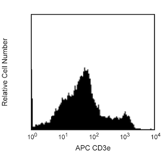
BD® CompBeads can be used as surrogates to assess fluorescence spillover (compensation). When fluorochrome conjugated antibodies are bound to BD® CompBeads, they have spectral properties very similar to cells. However, for some fluorochromes there can be small differences in spectral emissions compared to cells, resulting in spillover values that differ when compared to biological controls. It is strongly recommended that when using a reagent for the first time, users compare the spillover on cells and BD® CompBeads to ensure that BD® CompBeads are appropriate for your specific cellular application.

Product Notices

  1. Please refer to www.bdbiosciences.com/us/s/resources for technical protocols.
  2. Since applications vary, each investigator should titrate the reagent to obtain optimal results.
  3. An isotype control should be used at the same concentration as the antibody of interest.
  4. Caution: Sodium azide yields highly toxic hydrazoic acid under acidic conditions. Dilute azide compounds in running water before discarding to avoid accumulation of potentially explosive deposits in plumbing.
  5. For fluorochrome spectra and suitable instrument settings, please refer to our Multicolor Flow Cytometry web page at www.bdbiosciences.com/colors.
  6. Please refer to http://regdocs.bd.com to access safety data sheets (SDS).
  7. CF™ is a trademark of Biotium, Inc.
568546 Rev. 1
Antibody Details
Down Arrow Up Arrow
M5/114.15.2

The M5/114.15.2 monoclonal antibody recognizes a polymorphic determinant shared by the I-A[b], I-A[d], I-A[q], I-E[d], and I-E[k] (but not I-A[f], I-A[k], or I-A[s]) MHC class II alloantigens that can be expressed by B cells, dendritic cells, monocytes, macrophages and activated T cells. It also reacts with cells from mice of the H-2[p] and H-2[r] haplotypes, and it is non-reactive with cells from NOD (H-2[g7]) mice. Flow cytometric analysis indicates that the M5/114.15.2 and 2G9 monoclonal antibodies have comparable reactivity on cells from mice with I-A[b], I-A[d], I-A[g7], I-A[q], I-E[d], and I-E[k] alloantigens.

568546 Rev. 1
Format Details
Down Arrow Up Arrow
RY586
The BD Horizon RealYellow™ 586 (RY586) Dye is part of the BD family of yellow-green dyes. It is a small organic fluorochrome with an excitation maximum (Ex Max) at 565-nm and an emission maximum (Em Max) at 586-nm. Driven by BD innovation, RY586 can be used on both spectral and conventional cytometers and is designed to be excited by the Yellow-Green laser (561-nm) with minimal excitation by the 488-nm Blue laser. For conventional instruments equipped with a Yellow-Green laser (561-nm), RY586 can be used as an alternative to PE and we recommend using an optical filter centered near 586-nm (eg, a 586/15-nm bandpass filter). For spectral instruments equipped with a Yellow-Green laser (561-nm), it can be used in conjunction with PE. Compared to PE, RY586 is similar in brightness, minimal spillover into Blue detectors, and increased spillover into the 610/20-nm (PE-CF594) detector. Please ensure that your instrument configuration (lasers and optical filters) is appropriate for this dye.
altImg
RY586
Yellow-Green 561 nm
564 nm
586 nm
568546 Rev.1
Citations & References
Down Arrow Up Arrow
View product citations for antibody "568546" on CiteAb

Development References (7)

  1. Bhattacharya A, Dorf ME, Springer TA. A shared alloantigenic determinant on Ia antigens encoded by the I-A and I-E subregions: evidence for I region gene duplication. J Immunol. 1981; 127(6):2488-2495. (Immunogen: Immunoprecipitation). View Reference
  2. Ernst DN, McQuitty DN, Weigle WO, Hobbs MV. Expression of membrane activation antigens on murine B lymphocytes stimulated with lipopolysaccharide. Cell Immunol. 1988; 114(1):161-173. (Clone-specific: Flow cytometry). View Reference
  3. Guo MW, Watanabe T, Mori E, Mori T. Molecular structure and function of CD4 on murine egg plasma membrane. Zygote. 1995; 3(1):65-73. (Clone-specific: Blocking). View Reference
  4. Hattori M, Buse JB, Jackson RA, et al. The NOD mouse: recessive diabetogenic gene in the major histocompatibility complex. Science. 1986; 231(4739):733-735. (Clone-specific). View Reference
  5. Nelson AJ, Hosier S, Brady W, Linsley PS, Farr AG. Medullary thymic epithelium expresses a ligand for CTLA4 in situ and in vitro. J Immunol. 1998; 151(5):2453-2461. (Clone-specific: Blocking, Immunofluorescence, Immunohistochemistry). View Reference
  6. Viville S, Neefjes J, Lotteau V, et al. Mice lacking the MHC class II-associated invariant chain. Cell. 1993; 72(4):635-648. (Clone-specific: Flow cytometry, Immunofluorescence). View Reference
  7. Yamashita I, Nagata T, Tada T, Nakayama T. CD69 cell surface expression identifies developing thymocytes which audition for T cell antigen receptor-mediated positive selection. Int Immunol. 1993; 5(9):1139-1150. (Clone-specific: Blocking). View Reference
View All (7) View Less
568546 Rev. 1

Please refer to Support Documents for Quality Certificates


Global - Refer to manufacturer's instructions for use and related User Manuals and Technical data sheets before using this products as described


Comparisons, where applicable, are made against older BD Technology, manual methods or are general performance claims.  Comparisons are not made against non-BD technologies, unless otherwise noted.

For Research Use Only. Not for use in diagnostic or therapeutic procedures.