Preparation And Storage
Recommended Assay Procedures
BD® CompBeads can be used as surrogates to assess fluorescence spillover (compensation). When fluorochrome conjugated antibodies are bound to BD® CompBeads, they have spectral properties very similar to cells. However, for some fluorochromes there can be small differences in spectral emissions compared to cells, resulting in spillover values that differ when compared to biological controls. It is strongly recommended that when using a reagent for the first time, users compare the spillover on cells and BD® CompBeads to ensure that BD® CompBeads are appropriate for your specific cellular application.
Product Notices
- When using high concentrations of antibody, background binding of this dye to erythroid fragments produced by ammonium chloride-based lysis, such as with BD Pharm Lyse™ Lysing Buffer (Cat. No. 555899), has been observed when the antibody conjugate was present during the lysis procedure. This may cause nonspecific staining of target cells, such as leukocytes, which have bound the resulting erythroid fragments. This background can be mitigated by any of the following: titrating the antibody conjugate to a lower concentration, fixing samples with formaldehyde, or removing erythrocytes before staining (eg, gradient centrifugation or pre-lysis with wash). This background has not been observed when cells were lysed with BD FACS™ Lysing Solution (Cat. No. 349202) after staining.
- Please refer to www.bdbiosciences.com/us/s/resources for technical protocols.
- Since applications vary, each investigator should titrate the reagent to obtain optimal results.
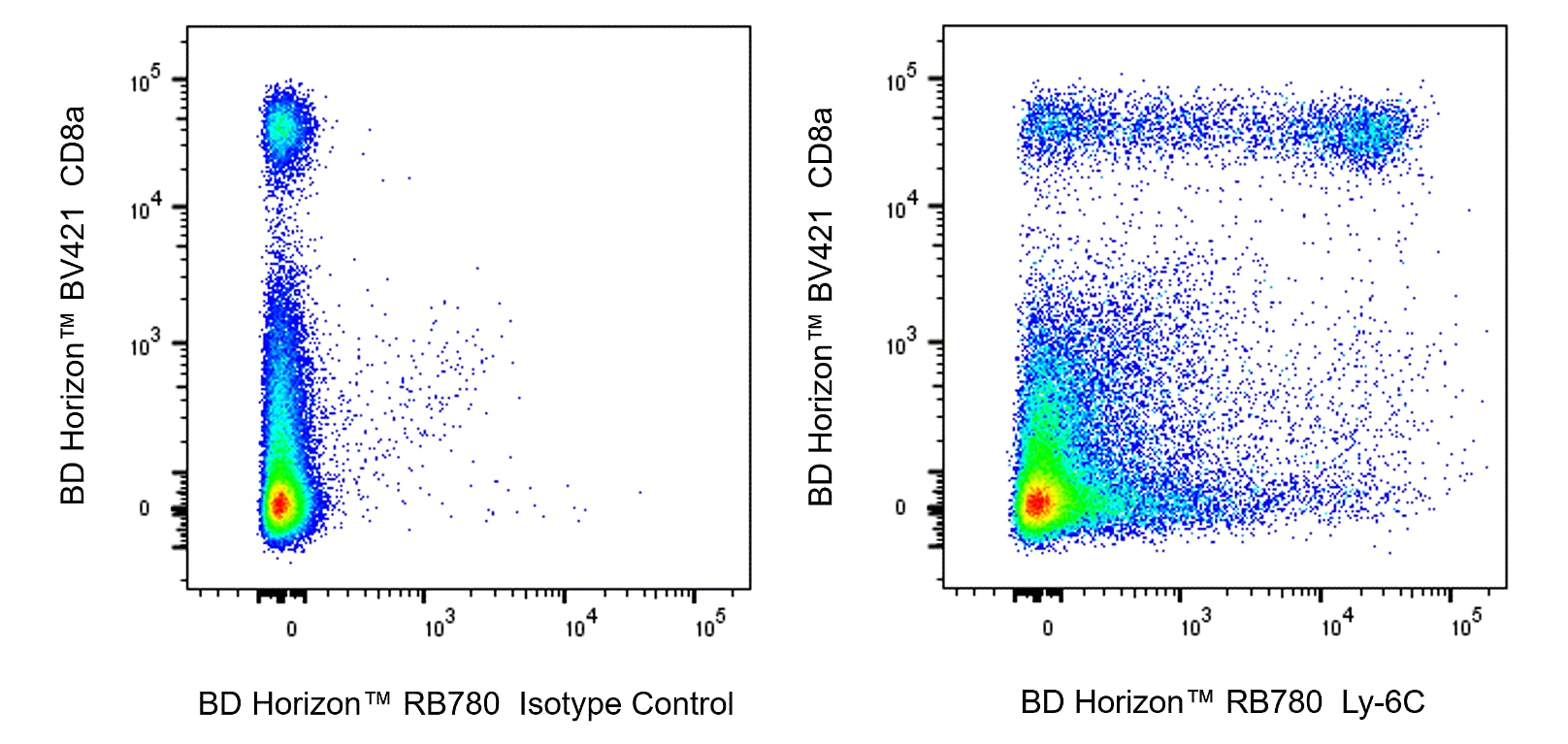
- An isotype control should be used at the same concentration as the antibody of interest.
- Please observe the following precautions: Absorption of visible light can significantly alter the energy transfer occurring in any tandem fluorochrome conjugate; therefore, we recommend that special precautions be taken (such as wrapping vials, tubes, or racks in aluminum foil) to prevent exposure of conjugated reagents, including cells stained with those reagents, to room illumination.
- Caution: Sodium azide yields highly toxic hydrazoic acid under acidic conditions. Dilute azide compounds in running water before discarding to avoid accumulation of potentially explosive deposits in plumbing.
- For fluorochrome spectra and suitable instrument settings, please refer to our Multicolor Flow Cytometry web page at www.bdbiosciences.com/colors.
- Please refer to http://regdocs.bd.com to access safety data sheets (SDS).
- Cy is a trademark of Global Life Sciences Solutions Germany GmbH or an affiliate doing business as Cytiva.
Companion Products


.png?imwidth=320)


The AL-21 monoclonal antibody specifically binds to a non-polymorphic determinant of Ly-6C, a 14-17 kDa GPI-linked cell-surface antigen found on some monocyte/macrophage populations, granulocytes, endothelial cells, plasma cells, and thymocyte, NK-cell, and T-subsets. Mice with the Ly-6.2 alloantigen (eg, AKR, C57BL, C57BR, C57L, C58, DBA/2, PL, SJL, SWR, 129) have subsets of CD8+ and CD4+ Ly-6C+ T cells, while Ly-6.1 strains (eg, A, BALB/c, CBA, C3H/He, DBA/1, NZB) have only CD8+ Ly-6C+ T cells. Upregulation of Ly-6C expression on CD8+ T cells by interferons α and β and poly (I:C) has been described, and Ly-6C is a memory marker on CD8+ T cells.

Development References (7)
-
Cerwenka A, Carter LL, Reome JB, Swain SL, Dutton RW. In vivo persistence of CD8 polarized T cell subsets producing type 1 or type 2 cytokines. J Immunol. 1998; 161(1):97-105. (Biology). View Reference
-
Jutila DB, Kurk S, Jutila MA.. Differences in the expression of Ly-6C on neutrophils and monocytes following PI-PLC hydrolysis and cellular activation.. Immunol Lett. 1994 ; 41(1):49-57. (Biology). View Reference
-
Jutila MA, Kroese FG, Jutila KL, et al. Ly-6C is a monocyte/macrophage and endothelial cell differentiation antigen regulated by interferon-gamma. Eur J Immunol. 1988; 18(11):1819-1826. (Biology). View Reference
-
Sato N, Yahata T, Santa K. Functional characterization of NK1.1 + Ly-6C+ cells. Immunol Lett. 1996; 1(54):1-5-9. (Clone-specific: Flow cytometry, Fluorescence activated cell sorting). View Reference
-
Takahama Y, Sharrow SO, Singer A. Expression of an unusual T cell receptor (TCR)-V beta repertoire by Ly-6C+ subpopulations of CD4+ and/or CD8+ thymocytes. Evidence for a developmental relationship between Ly-6C+ thymocytes and CD4-CD8-TCR-alpha beta+ thymocytes. J Immunol. 1991; 147(9):2883-2891. (Clone-specific: Flow cytometry). View Reference
-
Tough DF, Borrow P, Sprent J. Induction of bystander T cell proliferation by viruses and type I interferon in vivo. Science. 1996; 272(5270):1947-1950. (Biology). View Reference
-
Wrammert J, Källberg E, Agace WW, Leanderson T. Ly6C expression differentiates plasma cells from other B cell subsets in mice. Eur J Immunol. 2002; 32(1):97-103. (Clone-specific: Flow cytometry). View Reference
Please refer to Support Documents for Quality Certificates
Global - Refer to manufacturer's instructions for use and related User Manuals and Technical data sheets before using this products as described
Comparisons, where applicable, are made against older BD Technology, manual methods or are general performance claims. Comparisons are not made against non-BD technologies, unless otherwise noted.
For Research Use Only. Not for use in diagnostic or therapeutic procedures.