Preparation And Storage
Recommended Assay Procedures
BD® CompBeads can be used as surrogates to assess fluorescence spillover (compensation). When fluorochrome conjugated antibodies are bound to BD® CompBeads, they have spectral properties very similar to cells. However, for some fluorochromes there can be small differences in spectral emissions compared to cells, resulting in spillover values that differ when compared to biological controls. It is strongly recommended that when using a reagent for the first time, users compare the spillover on cells and BD® CompBeads to ensure that BD® CompBeads are appropriate for your specific cellular application.
Product Notices
- Please refer to www.bdbiosciences.com/us/s/resources for technical protocols.
- Please refer to http://regdocs.bd.com to access safety data sheets (SDS).
- For U.S. patents that may apply, see bd.com/patents.
- Caution: Sodium azide yields highly toxic hydrazoic acid under acidic conditions. Dilute azide compounds in running water before discarding to avoid accumulation of potentially explosive deposits in plumbing.
- Since applications vary, each investigator should titrate the reagent to obtain optimal results.
- The production process underwent stringent testing and validation to assure that it generates a high-quality conjugate with consistent performance and specific binding activity. However, verification testing has not been performed on all conjugate lots.
- Human donor specific background has been observed in relation to the presence of anti-polyethylene glycol (PEG) antibodies, developed as a result of certain vaccines containing PEG, including some COVID-19 vaccines. We recommend use of BD Horizon Brilliant™ Stain Buffer in your experiments to help mitigate potential background. For more information visit https://www.bdbiosciences.com/en-us/support/product-notices.
- When using high concentrations of antibody, background binding of this dye to erythroid fragments produced by ammonium chloride-based lysis, such as with BD Pharm Lyse™ Lysing Buffer (Cat. No. 555899), has been observed when the antibody conjugate was present during the lysis procedure. This may cause nonspecific staining of target cells, such as leukocytes, which have bound the resulting erythroid fragments. This background can be mitigated by any of the following: titrating the antibody conjugate to a lower concentration, fixing samples with formaldehyde, or removing erythrocytes before staining (eg, gradient centrifugation or pre-lysis with wash). This background has not been observed when cells were lysed with BD FACS™ Lysing Solution (Cat. No. 349202) after staining.
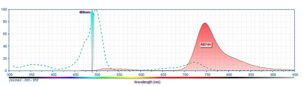
- For fluorochrome spectra and suitable instrument settings, please refer to our Multicolor Flow Cytometry web page at www.bdbiosciences.com/colors.
- An isotype control should be used at the same concentration as the antibody of interest.
- Please observe the following precautions: We recommend that special precautions be taken (such as wrapping vials, tubes, or racks in aluminum foil) to protect exposure of conjugated reagents, including cells stained with those reagents, to any room illumination. Absorption of visible light can significantly affect the emission spectra and quantum yield of tandem fluorochrome conjugates.
Companion Products


The F2.67 monoclonal antibody specifically recognizes the variable gamma 7 region of the γ subunit of the mouse γδ T cell receptor for antigen, TCR Vγ7 (using the Heilig and Tonegawa nomenclature for mouse TCR γ and δ chains). TCR Vγ7 is encoded by the Trgv7 (T cell receptor gamma, variable 7) gene element. TCR Vγ7 is expressed by a subset of TCR γδ+ thymocytes in the late fetal and adult thymus and by γδ T cells in peripheral lymphoid tissues. TCR Vγ7+ γδ T cells predominate in intestinal epithelial tissue which contains a large proportion of these γδ T cells derived from extrathymic generation. Proteins encoded by Btnl1 (butyrophilin-like 1) and Btnl6 (butyrophilin-like 6) are expressed by intestinal epithelial cells. These butyrophilin-like molecules can reportedly shape the TCR-dependent development and function of TCR Vg7+ γδ T cells within the gut. TCR Vγ7+ γδ T cells help maintain the integrity of the intestinal mucosa guarding against cellular stress or damage caused by inflammation, transformation, or infection. The F2.67 antibody is useful for TCR Vγ7+ thymocyte and γδ T cell separations and analyzing TCR Vγ repertoires expressed by thymocytes, peripheral T cells, and T cell hybridomas in developmental and other experimental model systems.
Development References (10)
-
Cossarizza A, Chang HD, Radbruch A, et al. Guidelines for the use of flow cytometry and cell sorting in immunological studies (second edition).. Eur J Immunol. 2019; 49(10):1457-1973. (Clone-specific: Flow cytometry). View Reference
-
Dalton JE, Cruickshank SM, Egan CE, et al. Intraepithelial gammadelta+ lymphocytes maintain the integrity of intestinal epithelial tight junctions in response to infection.. Gastroenterology. 2006; 131(3):818-29. (Clone-specific: Flow cytometry). View Reference
-
Garman RD, Doherty PJ, Raulet DH. Diversity, rearrangement, and expression of murine T cell gamma genes.. Cell. 1986; 45(5):733-42. (Biology). View Reference
-
Heilig JS, Tonegawa S. Diversity of murine gamma genes and expression in fetal and adult T lymphocytes.. Nature. 322(6082):836-40. (Biology: Flow cytometry). View Reference
-
Kashani E, Föhse L, Raha S, et al. A clonotypic Vγ4Jγ1/Vδ5Dδ2Jδ1 innate γδ T-cell population restricted to the CCR6⁺CD27⁻ subset.. Nat Commun. 2015; 6:6477. (Clone-specific: Flow cytometry). View Reference
-
Monin L, Ushakov DS, Arnesen H, et al. γδ T cells compose a developmentally regulated intrauterine population and protect against vaginal candidiasis.. Mucosal Immunol. 2020; 13(6):969-981. (Clone-specific: Flow cytometry). View Reference
-
Pereira P, Boucontet L. Rates of recombination and chain pair biases greatly influence the primary gammadelta TCR repertoire in the thymus of adult mice.. J Immunol. 2004; 173(5):3261-70. (Clone-specific: Flow cytometry). View Reference
-
Pereira P, Hermitte V, Lembezat MP, Boucontet L, Azuara V, Grigoriadou K. Developmentally regulated and lineage-specific rearrangement of T cell receptor Valpha/delta gene segments.. Eur J Immunol. 2000; 30(7):1988-97. (Immunogen: Flow cytometry). View Reference
-
Sell S, Dietz M, Schneider A, Holtappels R, Mach M, Winkler TH. Control of murine cytomegalovirus infection by γδ T cells.. PLoS Pathog. 2015; 11(2):e1004481. (Clone-specific: Flow cytometry). View Reference
-
Zeng W, O'Brien RL, Born WK, Huang Y. Characterization of Mouse γδ T Cell Subsets in the Setting of Type-2 Immunity.. Methods Mol Biol. 2018; 1799:135-151. (Clone-specific: Flow cytometry). View Reference
Please refer to Support Documents for Quality Certificates
Global - Refer to manufacturer's instructions for use and related User Manuals and Technical data sheets before using this products as described
Comparisons, where applicable, are made against older BD Technology, manual methods or are general performance claims. Comparisons are not made against non-BD technologies, unless otherwise noted.
For Research Use Only. Not for use in diagnostic or therapeutic procedures.