Preparation And Storage
Recommended Assay Procedures
BD® CompBeads can be used as surrogates to assess fluorescence spillover (compensation). When fluorochrome conjugated antibodies are bound to BD® CompBeads, they have spectral properties very similar to cells. However, for some fluorochromes there can be small differences in spectral emissions compared to cells, resulting in spillover values that differ when compared to biological controls. It is strongly recommended that when using a reagent for the first time, users compare the spillover on cells and BD® CompBeads to ensure that BD® CompBeads are appropriate for your specific cellular application.
Product Notices
- Please refer to www.bdbiosciences.com/us/s/resources for technical protocols.
- Please refer to http://regdocs.bd.com to access safety data sheets (SDS).
- For U.S. patents that may apply, see bd.com/patents.
- Caution: Sodium azide yields highly toxic hydrazoic acid under acidic conditions. Dilute azide compounds in running water before discarding to avoid accumulation of potentially explosive deposits in plumbing.
- Since applications vary, each investigator should titrate the reagent to obtain optimal results.
- The production process underwent stringent testing and validation to assure that it generates a high-quality conjugate with consistent performance and specific binding activity. However, verification testing has not been performed on all conjugate lots.
- Human donor specific background has been observed in relation to the presence of anti-polyethylene glycol (PEG) antibodies, developed as a result of certain vaccines containing PEG, including some COVID-19 vaccines. We recommend use of BD Horizon Brilliant™ Stain Buffer in your experiments to help mitigate potential background. For more information visit https://www.bdbiosciences.com/en-us/support/product-notices.
- When using high concentrations of antibody, background binding of this dye to erythroid fragments produced by ammonium chloride-based lysis, such as with BD Pharm Lyse™ Lysing Buffer (Cat. No. 555899), has been observed when the antibody conjugate was present during the lysis procedure. This may cause nonspecific staining of target cells, such as leukocytes, which have bound the resulting erythroid fragments. This background can be mitigated by any of the following: titrating the antibody conjugate to a lower concentration, fixing samples with formaldehyde, or removing erythrocytes before staining (eg, gradient centrifugation or pre-lysis with wash). This background has not been observed when cells were lysed with BD FACS™ Lysing Solution (Cat. No. 349202) after staining.
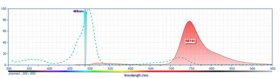
- For fluorochrome spectra and suitable instrument settings, please refer to our Multicolor Flow Cytometry web page at www.bdbiosciences.com/colors.
- An isotype control should be used at the same concentration as the antibody of interest.
- Please observe the following precautions: We recommend that special precautions be taken (such as wrapping vials, tubes, or racks in aluminum foil) to protect exposure of conjugated reagents, including cells stained with those reagents, to any room illumination. Absorption of visible light can significantly affect the emission spectra and quantum yield of tandem fluorochrome conjugates.
Companion Products


The A3 monoclonal antibody specifically binds to CD148. CD148 is a receptor-type tyrosine phosphatase that belongs to the receptor class 3 subfamily of the protein-tyrosine phosphatase family. CD148 is also known as Density-enhanced phosphatase 1 (DEP-1), Human protein tyrosine phosphatase- η (HPTP-η/HPTP-eta), or Protein tyrosine phosphatase receptor type J (PTPRJ). CD148 is a highly glycosylated type I transmembrane protein that is widely expressed on different cell types including monocytes, granulocytes, dendritic cells, thymocytes, T cells, B cells, NK cells, fibroblasts, and platelets. As a protein tyrosine phosphatase with receptor function, CD148 plays a role in regulating the signaling activities of various phosphorylated cellular proteins including the PDGF Receptor, catenins and Src family kinases. In the immune system, CD148 plays a role in regulating the activities of different cell types. For example, upon T cell activation, CD148 expression is upregulated by T cells. CD148 can subsequently dephosphorylate LAT and PLC-γ signaling proteins and thereby downregulate T cell signaling responses. Immobilized A3 antibody can reportedly augment the proliferation of anti-CD3 antibody-stimulated peripheral blood T cells in culture.
Development References (4)
-
Baker JE, Majeti R, Tangye SG, Weiss A. Protein tyrosine phosphatase CD148-mediated inhibition of T-cell receptor signal transduction is associated with reduced LAT and phospholipase Cgamma1 phosphorylation. Mol Cell Biol. 2001; 21(7):2393-2403. (Clone-specific: Flow cytometry). View Reference
-
Scraven B, Hegen M, Autschbach F, Gaya A, Schwarz C, Meuer SC. CD148 (p260 phosphatase) Workshop Panel report. In: Kishimoto T. Tadamitsu Kishimoto .. et al., ed. Leucocyte typing VI : white cell differentiation antigens : proceedings of the sixth international workshop and conference held in Kobe, Japan, 10-14 November 1996. New York: Garland Pub.; 1997:576-580.
-
Tangye SG, Phillips JH, Lanier LL, de Vries JE, Aversa G. CD148: a receptor-type protein tyrosine phosphatase involved in the regulation of human T cell activation. J Immunol. 1998; 161(7):9249-9255. (Immunogen: (Co)-stimulation, Flow cytometry, Functional assay). View Reference
-
Whiteford JR, Xian X, Chaussade C, Vanhaesebroeck B, Nourshargh S, Couchman JR. Syndecan-2 is a novel ligand for the protein tyrosine phosphatase receptor CD148. Mol Cell Biol. 2011; 22(19):3609-3624. (Clone-specific: Flow cytometry, Functional assay, Western blot). View Reference
Please refer to Support Documents for Quality Certificates
Global - Refer to manufacturer's instructions for use and related User Manuals and Technical data sheets before using this products as described
Comparisons, where applicable, are made against older BD Technology, manual methods or are general performance claims. Comparisons are not made against non-BD technologies, unless otherwise noted.
For Research Use Only. Not for use in diagnostic or therapeutic procedures.