Preparation And Storage
Recommended Assay Procedures
BD® CompBeads can be used as surrogates to assess fluorescence spillover (compensation). When fluorochrome conjugated antibodies are bound to BD® CompBeads, they have spectral properties very similar to cells. However, for some fluorochromes there can be small differences in spectral emissions compared to cells, resulting in spillover values that differ when compared to biological controls. It is strongly recommended that when using a reagent for the first time, users compare the spillover on cells and BD® CompBeads to ensure that BD® CompBeads are appropriate for your specific cellular application.
Product Notices
- Please refer to www.bdbiosciences.com/us/s/resources for technical protocols.
- Please observe the following precautions: Absorption of visible light can significantly alter the energy transfer occurring in any tandem fluorochrome conjugate; therefore, we recommend that special precautions be taken (such as wrapping vials, tubes, or racks in aluminum foil) to prevent exposure of conjugated reagents, including cells stained with those reagents, to room illumination.
- Caution: Sodium azide yields highly toxic hydrazoic acid under acidic conditions. Dilute azide compounds in running water before discarding to avoid accumulation of potentially explosive deposits in plumbing.
- This reagent has been pre-diluted for use at the recommended Volume per Test. We typically use 1 × 10^6 cells in a 100-µl experimental sample (a test).
- For fluorochrome spectra and suitable instrument settings, please refer to our Multicolor Flow Cytometry web page at www.bdbiosciences.com/colors.
- An isotype control should be used at the same concentration as the antibody of interest.
- Please refer to http://regdocs.bd.com to access safety data sheets (SDS).
- Species cross-reactivity detected in product development may not have been confirmed on every format and/or application.
- Human donor specific background has been observed in relation to the presence of anti-polyethylene glycol (PEG) antibodies, developed as a result of certain vaccines containing PEG, including some COVID-19 vaccines. We recommend use of BD Horizon Brilliant™ Stain Buffer in your experiments to help mitigate potential background. For more information visit https://www.bdbiosciences.com/en-us/support/product-notices.
- For U.S. patents that may apply, see bd.com/patents.
Companion Products


.png?imwidth=320)
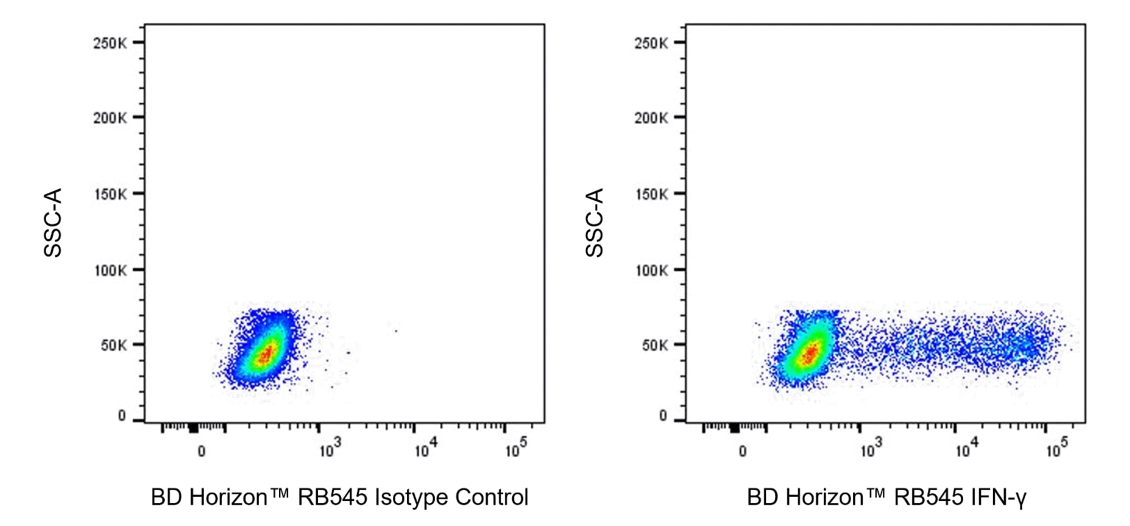
The B27 monoclonal antibody specifically binds to human interferon-γ (IFN-γ), a 14-18 kDa glycoprotein containing 143 amino acid residues. IFN-γ is a potent multifunctional cytokine produced by several activated cell types including NK, NKT, CD4+TCRαβ+, CD8+TCRαβ+, and TCRγδ+ T cells. IFN-γ exerts its biological effects through specific binding to the high-affinity IFN-γ receptor complex comprised of IFN-γRα (CD119) and IFN-γRβ subunits. In addition to its antiviral effects, IFN-γ upregulates a number of lymphoid cell functions including the antimicrobial and anti-tumor responses of macrophages, NK cells, and neutrophils. In addition, IFN-γ influences the regulation of proliferation, differentiation, and effector responses of B cell and T cell subsets. These influences can involve IFN-γ's capacity to boost MHC class I and II expression by antigen-presenting cells as well as direct effects on B cells and T cells themselves. B27 is a neutralizing antibody. The use of B27 antibody for epitope mapping of human IFN-γ has been described. The B27 antibody has been reported not to bind to denatured IFN-γ.

Development References (7)
-
Abrams JS, Roncarolo MG, Yssel H, Andersson U, Gleich GJ, Silver JE. Strategies of anti-cytokine monoclonal antibody development: immunoassay of IL-10 and IL-5 in clinical samples. Immunol Rev. 1992; 127:5-24. (Clone-specific: ELISA, Neutralization). View Reference
-
Draghi M, Yawata N, Gleimer M, Yawata M, Valiante NM, Parham P. Single-cell analysis of the human NK cell response to missing self and its inhibition by HLA class I.. Blood. 2005; 105(5):2028-35. (Clone-specific: Flow cytometry). View Reference
-
Favre C, Wijdenes J, Cabrillat H, Djossou O, Banchereau J, de Vries JE. Epitope mapping of recombinant human gamma interferon using monoclonal antibodies. Mol Immunol. 1989; 26(1):17-25. (Clone-specific: Flow cytometry, Immunoprecipitation, Neutralization). View Reference
-
Prussin C, Metcalfe DD. Detection of intracytoplasmic cytokine using flow cytometry and directly conjugated anti-cytokine antibodies. J Immunol Methods. 1995; 188(1):117-128. (Methodology: Flow cytometry, IC/FCM Block). View Reference
-
Rosato PC, Wijeyesinghe S, Stolley JM, et al. Virus-specific memory T cells populate tumors and can be repurposed for tumor immunotherapy.. Nat Commun. 2019; 10(1):567. (Clone-specific: Flow cytometry). View Reference
-
Satooka H, Ishigaki H, Todo K, et al. Characterization of tumour-infiltrating lymphocytes in a tumour rejection cynomolgus macaque model. Scientific reports. 2020; 10(1):8414. (Clone-specific: Flow cytometry). View Reference
-
Trotta E, Bessette PH, Silveria SL, et al. A human anti-IL-2 antibody that potentiates regulatory T cells by a structure-based mechanism.. Nat Med. 2018; 24(7):1005-1014. (Clone-specific: Flow cytometry). View Reference
Please refer to Support Documents for Quality Certificates
Global - Refer to manufacturer's instructions for use and related User Manuals and Technical data sheets before using this products as described
Comparisons, where applicable, are made against older BD Technology, manual methods or are general performance claims. Comparisons are not made against non-BD technologies, unless otherwise noted.
For Research Use Only. Not for use in diagnostic or therapeutic procedures.