Preparation And Storage
Recommended Assay Procedures
BD® CompBeads can be used as surrogates to assess fluorescence spillover (compensation). When fluorochrome conjugated antibodies are bound to BD® CompBeads, they have spectral properties very similar to cells. However, for some fluorochromes there can be small differences in spectral emissions compared to cells, resulting in spillover values that differ when compared to biological controls. It is strongly recommended that when using a reagent for the first time, users compare the spillover on cells and BD® CompBeads to ensure that BD® CompBeads are appropriate for your specific cellular application.
Product Notices
- Please refer to www.bdbiosciences.com/us/s/resources for technical protocols.
- Since applications vary, each investigator should titrate the reagent to obtain optimal results.
- An isotype control should be used at the same concentration as the antibody of interest.
- Caution: Sodium azide yields highly toxic hydrazoic acid under acidic conditions. Dilute azide compounds in running water before discarding to avoid accumulation of potentially explosive deposits in plumbing.
- Please refer to http://regdocs.bd.com to access safety data sheets (SDS).
- This product is provided under an Agreement between BIOTIUM and BD Biosciences. This product, and only in the amount purchased by buyer, may be used solely for buyer’s own internal research, in a manner consistent with the accompanying product literature. No other right to use, sell or otherwise transfer (a) this product, or (b) its components is hereby granted expressly, by implication or by estoppel. This product is for research use only. Diagnostic uses require a separate license from Biotium, Inc. For information on purchasing a license to this product including for purposes other than research, contact Biotium, Inc., 3159 Corporate Place, Hayward, CA 94545, Tel: (510) 265-1027. Fax: (510) 265-1352. Email: btinfo@biotium.com.
- Alexa Fluor™ is a trademark of Life Technologies Corporation.
- For U.S. patents that may apply, see bd.com/patents.
Companion Products


.png?imwidth=320)
.png?imwidth=320)

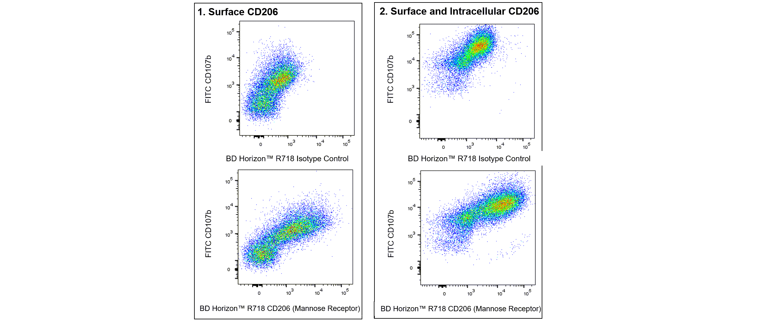
The Y17-505 monoclonal antibody specifically binds to CD206 which is also known as the Macrophage mannose receptor (MMR, MR) or Mannose receptor, C type 1 (Mrc1). CD206 is a type I transmembrane glycoprotein of approximately 175 kDa that belongs to the C-type lectin superfamily. It is expressed at the cell surface and intracellularly by macrophages, Langerhans cells, dendritic cells, and endothelial cells within hepatic and lymphoid tissues. This pattern recognition receptor binds to endogenous and microbial glycoconjugates containing mannose, fucose, or N-acetylglucosamine through its C-type lectin-like carbohydrate recognition domains (CRD). CD206 also contains a cysteine-rich domain that enables binding to sulfated carbohydrate antigens. This receptor enables macrophages and other specialized cells to maintain tissue homeostasis as well as to internalize microbes or their components by phagocytosis or endocytosis. CD206 thus plays important roles in mediating innate immunity, eg, enabling phagocytosis, as well as in processing and presenting antigens for the generation and expression of adaptive immunity. Moreover, CD206 has been associated with leucocyte homing and cancer cell metastasis.

Development References (6)
-
Akbarshahi H, Menzel M, Posaric Bauden M, Rosendahl A, Andersson R. Enrichment of murine CD68+ CCR2+ and CD68+ CD206+ lung macrophages in acute pancreatitis-associated acute lung injury. PLoS ONE. 2012; 7(10):e42654. (Biology). View Reference
-
Burgdorf S, Lukacs-Kornek V, Kurts C. The mannose receptor mediates uptake of soluble but not of cell-associated antigen for cross-presentation. J Immunol. 2006; 176(11):6770-6776. (Biology). View Reference
-
Marttila-Ichihara F, Turja R, Miiluniemi M, et al. Macrophage mannose receptor on lymphatics controls cell trafficking. Blood. 2008; 112(1):64-72. (Biology). View Reference
-
McKenzie EJ, Taylor PR, Stillion RJ, et al. J Immunol. 2007; 178(8):4975-4983. (Biology). View Reference
-
Rybalko V, Hsieh PL, Merscham-Banda M, Suggs LJ, Farrar RP. The Development of Macrophage-Mediated Cell Therapy to Improve Skeletal Muscle Function after Injury.. PLoS One. 2015; 10(12):e0145550. (Biology). View Reference
-
Zamze S, Martinez-Pomares L, Jones H, et al. Recognition of bacterial capsular polysaccharides and lipopolysaccharides by the macrophage mannose receptor. J Biol Chem. 2002; 277(44):41613-41623. (Biology). View Reference
Please refer to Support Documents for Quality Certificates
Global - Refer to manufacturer's instructions for use and related User Manuals and Technical data sheets before using this products as described
Comparisons, where applicable, are made against older BD Technology, manual methods or are general performance claims. Comparisons are not made against non-BD technologies, unless otherwise noted.
For Research Use Only. Not for use in diagnostic or therapeutic procedures.