Skip to main content Skip to navigation
FITC Mouse Anti-Human HLA-A3
Multiparameter flow cytometric analysis of HLA-A3 expression on Human peripheral blood leukocyte populations. Human whole blood was stained with either FITC Mouse IgG2a, κ Isotype Control (Cat. No. 553456; Left Plot) or FITC Mouse Anti-Human HLA-A3 antibody (Cat. No. 570461/570463; Right Plot) at 0.5 µg/test. Erythrocytes were lysed with BD Pharm Lyse™ Lysing Buffer (Cat. No. 555899). The bivariate pseudocolor density plot showing the correlated expression of HLA-A3 (or Ig Isotype control staining) versus side light-scatter (SSC-A) signals was derived from gated events with the forward and side light-scatter characteristics of viable leukocyte populations. Flow cytometric analysis was performed using a BD LSRFortessa™ X-20 Cell Analyzer System and  FlowJo™ Software. Data shown on this Technical Data Sheet are not lot specific.
FITC Mouse Anti-Human HLA-A3
Flow cytometric analysis of HLA-A3 expression on viable Jurkat cells. Jurkat cells were stained with FITC Mouse IgG2a, κ Isotype Control (dashed line histogram) or FITC Mouse Anti-Human HLA-A3 antibody (solid line histogram) at 0.5 µg/test. DAPI (4',6-Diamidino-2-Phenylindole, Dihydrochloride) Solution (Cat. No. 564907) was added to cells right before analysis. The fluorescence histogram showing HLA-A3 expression (or Ig Isotype control staining) was derived from gated events with the forward and side light-scatter characteristics of viable (DAPI-negative) cells. Flow cytometric analysis was performed using a BD LSRFortessa™ X-20 Cell Analyzer System and FlowJo™ Software. Data shown on this Technical Data Sheet are not lot specific.
Multiparameter flow cytometric analysis of HLA-A3 expression on Human peripheral blood leukocyte populations. Human whole blood was stained with either FITC Mouse IgG2a, κ Isotype Control (Cat. No. 553456; Left Plot) or FITC Mouse Anti-Human HLA-A3 antibody (Cat. No. 570461/570463; Right Plot) at 0.5 µg/test. Erythrocytes were lysed with BD Pharm Lyse™ Lysing Buffer (Cat. No. 555899). The bivariate pseudocolor density plot showing the correlated expression of HLA-A3 (or Ig Isotype control staining) versus side light-scatter (SSC-A) signals was derived from gated events with the forward and side light-scatter characteristics of viable leukocyte populations. Flow cytometric analysis was performed using a BD LSRFortessa™ X-20 Cell Analyzer System and  FlowJo™ Software. Data shown on this Technical Data Sheet are not lot specific.
Flow cytometric analysis of HLA-A3 expression on viable Jurkat cells. Jurkat cells were stained with FITC Mouse IgG2a, κ Isotype Control (dashed line histogram) or FITC Mouse Anti-Human HLA-A3 antibody (solid line histogram) at 0.5 µg/test. DAPI (4',6-Diamidino-2-Phenylindole, Dihydrochloride) Solution (Cat. No. 564907) was added to cells right before analysis. The fluorescence histogram showing HLA-A3 expression (or Ig Isotype control staining) was derived from gated events with the forward and side light-scatter characteristics of viable (DAPI-negative) cells. Flow cytometric analysis was performed using a BD LSRFortessa™ X-20 Cell Analyzer System and FlowJo™ Software. Data shown on this Technical Data Sheet are not lot specific.
Product Details
Down Arrow Up Arrow


BD Pharmingen™
HLA class I histocompatibility antigen, A-3 alpha chain; HLA Antigen A3
Human (QC Testing)
Mouse BALB/c IgG2a, κ
Human Lymphocytes from Mixed Lymphocyte Reaction
Flow cytometry (Routinely Tested)
0.5 mg/ml
3105
Aqueous buffered solution containing ≤0.09% sodium azide.
RUO


Preparation And Storage

The monoclonal antibody was purified from tissue culture supernatant or ascites by affinity chromatography. The antibody was conjugated to the dye under optimum conditions and unconjugated antibody and free dye were removed. Store undiluted at 4°C and protected from prolonged exposure to light. Do not freeze.

Recommended Assay Procedures

BD® CompBeads can be used as surrogates to assess fluorescence spillover (compensation).  When fluorochrome conjugated antibodies are bound to BD® CompBeads, they have spectral properties very similar to cells.  However, for some fluorochromes there can be small differences in spectral emissions compared to cells, resulting in spillover values that differ when compared to biological controls.  It is strongly recommended that when using a reagent for the first time, users compare the spillover on cell and BD® CompBeads to ensure that BD® CompBeads are appropriate for your specific cellular application.

Product Notices

  1. Please refer to www.bdbiosciences.com/us/s/resources for technical protocols.
  2. Since applications vary, each investigator should titrate the reagent to obtain optimal results.
  3. An isotype control should be used at the same concentration as the antibody of interest.
  4. Caution: Sodium azide yields highly toxic hydrazoic acid under acidic conditions. Dilute azide compounds in running water before discarding to avoid accumulation of potentially explosive deposits in plumbing.
  5. For fluorochrome spectra and suitable instrument settings, please refer to our Multicolor Flow Cytometry web page at www.bdbiosciences.com/colors.
  6. Please refer to http://regdocs.bd.com to access safety data sheets (SDS).
  7. For U.S. patents that may apply, see bd.com/patents.
570463 Rev. 1
Antibody Details
Down Arrow Up Arrow
GAP.A3

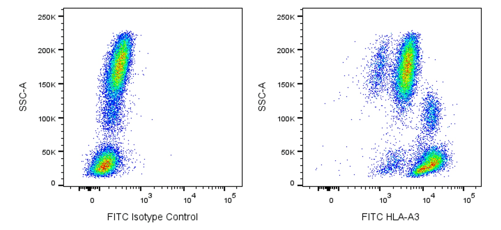
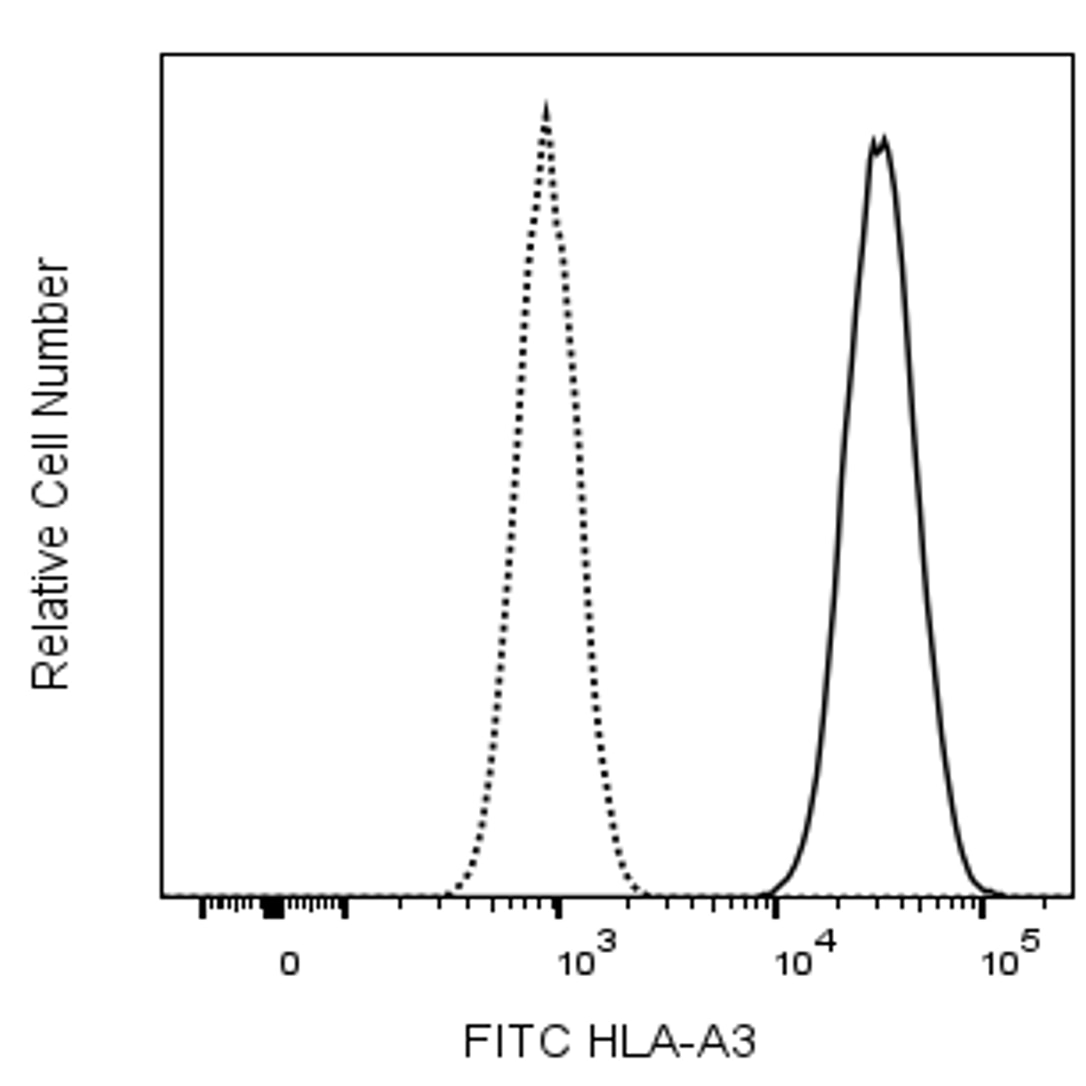
The GAP.A3 monoclonal antibody specifically recognizes the polymorphic HLA class I histocompatibility antigen, A-3 alpha chain (HLA-A3). This ~44 kDa human major histocompatibility complex (MHC) class I molecule noncovalently associates with the ~12 kDa monomorphic β2 microglobulin. Two major subtypes are encoded by HLA-A3, HLA-A3.1 and HLA-A3.2. HLA-A3 gene expression is more commonly detected in individuals from Europe and southern India. HLA-A3 is expressed on nearly all nucleated cells of HLA-A3-positive individuals. HLA-A3 functions in the presentation of antigens to CD8-positive T cells which may lead to the generation of HLA-A3-restricted cytotoxic T cells and memory T cells. When complexed with certain antigens, HLA-A3 can also bind to killer cell immunoglobulin-like receptors (KIR), such as KIR3DL2, and might regulate NK cell function.

Note: Since HLA-A3 expression varies between human populations, clone GAP.A3 staining can be donor-dependent. Based on in-house testing and current literature, individuals of European or Southern Indian descent more frequently express HLA-A3 than those of Asian descent. Data may differ between donors due to geographical variations of HLA-A3 expression.

570463 Rev. 1
Format Details
Down Arrow Up Arrow
FITC
Fluorescein (FITC) is part of the BD blue family of dyes. This is a small organic fluorochrome with an excitation maximum (Ex Max) at 494-nm and an emission maximum (Em Max) at 518-nm. FITC is designed to be excited by the Blue laser (488-nm) and detected using an optical filter centered near 520 nm (e.g., a 530/30-nm bandpass filter). Please ensure that your instrument’s configurations (lasers and optical filters) are appropriate for this dye.
altImg
FITC
Blue 488 nm
494 nm
518 nm
570463 Rev.1
Citations & References
Down Arrow Up Arrow
View product citations for antibody "570463" on CiteAb

Development References (7)

  1. Allen TM, Altfeld M, Yu XG, et al. Selection, transmission, and reversion of an antigen-processing cytotoxic T-lymphocyte escape mutation in human immunodeficiency virus type 1 infection.. J Virol. 2004; 78(13):7069-78. (Biology). View Reference
  2. Augusto DG, O'Connor GM, Lobo-Alves SC, et al. Pemphigus is associated with KIR3DL2 expression levels and provides evidence that KIR3DL2 may bind HLA-A3 and A11 in vivo.. Eur J Immunol. 2015; 45(7):2052-60. (Biology). View Reference
  3. Berger AE, Davis JE, Cresswell P. Monoclonal antibody to HLA-A3.. Hybridoma. 1982; 1(2):87-90. (Immunogen). View Reference
  4. Carr TM, Adair SJ, Fink MJ, Hogan KT. Immunological profiling of a panel of human ovarian cancer cell lines.. Cancer Immunol Immunother. 2008; 57(1):31-42. (Clone-specific). View Reference
  5. Hansasuta P, Dong T, Thananchai H, et al. Recognition of HLA-A3 and HLA-A11 by KIR3DL2 is peptide-specific.. Eur J Immunol. 2004; 34(6):1673-9. (Biology). View Reference
  6. Lalouel JM, Jorde LB. Idiopathic hemochromatosis: significance and implications of linkage and association to HLA.. Ann N Y Acad Sci. 1988; 526:34-46. (Biology). View Reference
  7. Vonderheide RH, Anderson KS, Hahn WC, Butler MO, Schultze JL, Nadler LM. Characterization of HLA-A3-restricted cytotoxic T lymphocytes reactive against the widely expressed tumor antigen telomerase.. Clin Cancer Res. 2001; 7(11):3343-8. (Biology). View Reference
View All (7) View Less
570463 Rev. 1

Please refer to Support Documents for Quality Certificates


Global - Refer to manufacturer's instructions for use and related User Manuals and Technical data sheets before using this products as described


Comparisons, where applicable, are made against older BD Technology, manual methods or are general performance claims.  Comparisons are not made against non-BD technologies, unless otherwise noted.

For Research Use Only. Not for use in diagnostic or therapeutic procedures.