
Preparation And Storage
Recommended Assay Procedures
BD® CompBeads can be used as surrogates to assess fluorescence spillover (compensation). When fluorochrome conjugated antibodies are bound to BD® CompBeads, they have spectral properties very similar to cells. However, for some fluorochromes there can be small differences in spectral emissions compared to cells, resulting in spillover values that differ when compared to biological controls. It is strongly recommended that when using a reagent for the first time, users compare the spillover on cells and BD® CompBeads to ensure that BD® CompBeads are appropriate for your specific cellular application.
For optimal and reproducible results, BD Horizon Brilliant™ Stain Buffer should be used anytime BD Horizon Brilliant dyes are used in a multicolor flow cytometry panel. Fluorescent dye interactions may cause staining artifacts which may affect data interpretation. The BD Horizon Brilliant Stain Buffer was designed to minimize these interactions. When BD Horizon Brilliant Stain Buffer is used in in the multicolor panel, it should also be used in the corresponding compensation controls for all dyes to achieve the most accurate compensation. For the most accurate compensation, compensation controls created with either cells or beads should be exposed to BD Horizon Brilliant Stain Buffer for the same length of time as the corresponding multicolor panel. More information can be found in the Technical Data Sheet of the BD Horizon Brilliant Stain Buffer (Cat. No. 563794/566349) or the BD Horizon Brilliant Stain Buffer Plus (Cat. No. 566385).
Product Notices
- Please refer to www.bdbiosciences.com/us/s/resources for technical protocols.
- Please observe the following precautions: Absorption of visible light can significantly alter the energy transfer occurring in any tandem fluorochrome conjugate; therefore, we recommend that special precautions be taken (such as wrapping vials, tubes, or racks in aluminum foil) to prevent exposure of conjugated reagents, including cells stained with those reagents, to room illumination.
- Caution: Sodium azide yields highly toxic hydrazoic acid under acidic conditions. Dilute azide compounds in running water before discarding to avoid accumulation of potentially explosive deposits in plumbing.
- This reagent has been pre-diluted for use at the recommended Volume per Test. We typically use 1 × 10^6 cells in a 100-µl experimental sample (a test).
- For fluorochrome spectra and suitable instrument settings, please refer to our Multicolor Flow Cytometry web page at www.bdbiosciences.com/colors.
- An isotype control should be used at the same concentration as the antibody of interest.
- CF™ is a trademark of Biotium, Inc.
- Although every effort is made to minimize the lot-to-lot variation in the efficiency of the fluorochrome energy transfer, differences in the residual emission from BD Horizon™ BV421 may be observed. Therefore, we recommend that individual compensation controls be performed for every BD Horizon™ BV605 conjugate.
- BD Horizon Brilliant Violet 605 is covered by one or more of the following US patents: 8,110,673; 8,158,444; 8,227,187; 8,455,613; 8,575,303; 8,354,239.
- BD Horizon Brilliant Stain Buffer is covered by one or more of the following US patents: 8,110,673; 8,158,444; 8,575,303; 8,354,239.
- Please refer to http://regdocs.bd.com to access safety data sheets (SDS).
- Human donor specific background has been observed in relation to the presence of anti-polyethylene glycol (PEG) antibodies, developed as a result of certain vaccines containing PEG, including some COVID-19 vaccines. We recommend use of BD Horizon Brilliant™ Stain Buffer in your experiments to help mitigate potential background. For more information visit https://www.bdbiosciences.com/en-us/support/product-notices.
- For U.S. patents that may apply, see bd.com/patents.

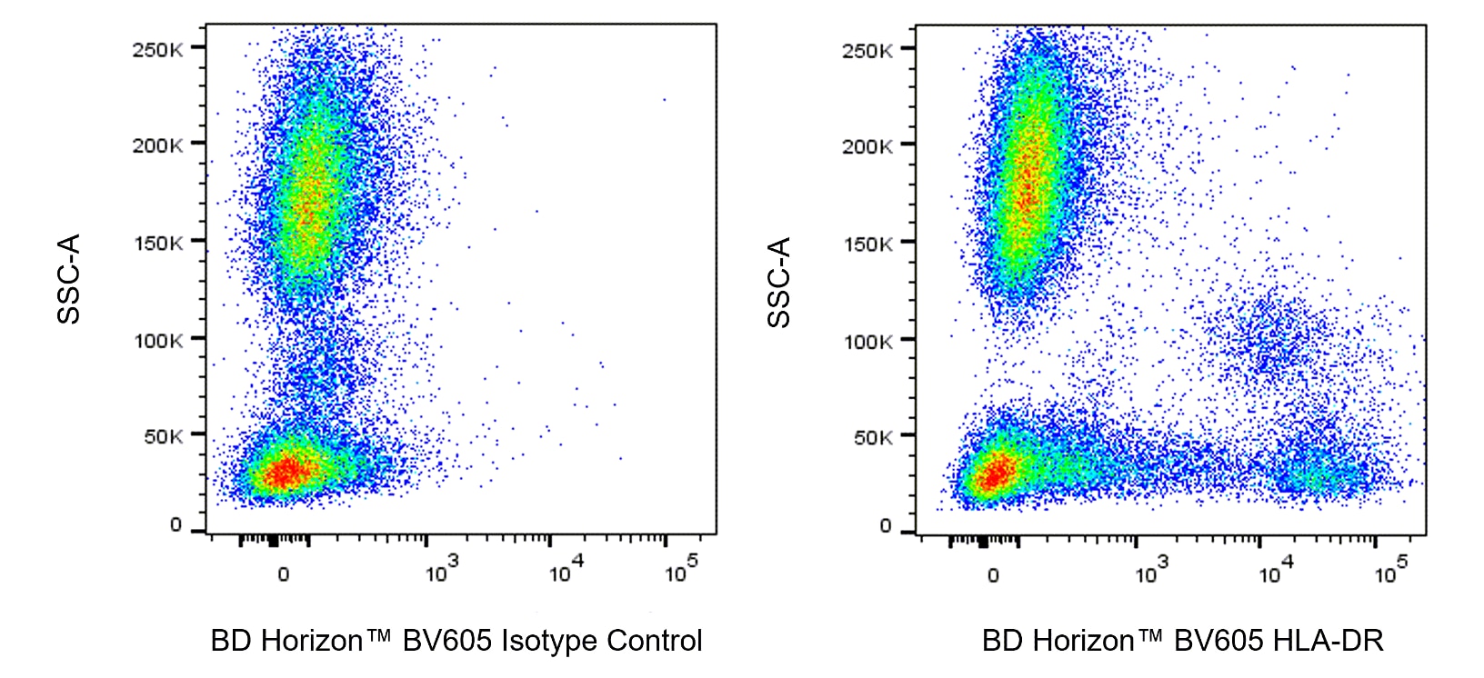
The L243 monoclonal antibody specifically binds to HLA-DR, a major histocompatibility complex (MHC) class II antigen. HLA-DR antigens are encoded by genes within the Human Leukocyte Antigen (HLA) Complex located on chromosome 6. HLA-DR is a transmembrane heterodimeric glycoprotein composed of an α chain (36 kDa) and a β subunit (27 kDa) expressed primarily on antigen presenting cells including B cells, dendritic cells, monocytes, macrophages, Langerhans cells, and thymic epithelial cells. HLA-DR is also expressed on activated T cells. This molecule plays a major role in mediating cellular interactions during antigen presentation to CD4-positive T cells.

Development References (12)
-
Autissier P, Soulas C, Burdo TH, Williams KC. Evaluation of a 12-color flow cytometry panel to study lymphocyte, monocyte, and dendritic cell subsets in humans.. Cytometry A. 2010; 77(5):410-9. (Clone-specific: Flow cytometry). View Reference
-
Brodsky FM. A matrix approach to human class II histocompatibility antigens: reactions of four monoclonal antibodies with the products of nine haplotypes.. Immunogenetics. 1984; 19(3):179-94. (Clone-specific: Blocking, Immunoprecipitation, Radioimmunoassay). View Reference
-
Charles N, Hardwick D, Daugas E, Illei GG, Rivera J. Basophils and the T helper 2 environment can promote the development of lupus nephritis.. Nat Med. 2010; 16(6):701-7. (Clone-specific: Flow cytometry). View Reference
-
Engleman EG, Warnke R, Fox RI, Dilley J, Benike CJ, Levy R. Studies of a human T lymphocyte antigen recognized by a monoclonal antibody.. Proc Natl Acad Sci USA. 1981; 78(3):1791-5. (Clone-specific: Immunohistochemistry). View Reference
-
Fujita H, Nograles KE, Kikuchi T, Gonzalez J, Carucci JA, Krueger JG. Human Langerhans cells induce distinct IL-22-producing CD4+ T cells lacking IL-17 production.. Proc Natl Acad Sci U S A. 2009; 106(51):21795-800. (Clone-specific: Flow cytometry). View Reference
-
Harper D, Gollackner B, Xu Y, et al. In vitro and in vivo investigation of a novel monoclonal antibody to plasma cells (W5 mAb).. Xenotransplantation. 2004; 11(1):78-90. (Clone-specific: Immunoprecipitation). View Reference
-
Lampson LA, Levy R. Two populations of Ia-like molecules on a human B cell line.. J Immunol. 1980; 125(1):293-9. (Immunogen: Blocking, Immunoprecipitation, Radioimmunoassay). View Reference
-
Ren Z, Wang J, Zhu W, et al. Spontaneous transformation of adult mesenchymal stem cells from cynomolgus macaques in vitro.. Exp Cell Res. 2011; 317(20):2950-7. (Clone-specific: Flow cytometry). View Reference
-
Stumptner-Cuvelette P, Morchoisne S, Dugast M, et al. HIV-1 Nef impairs MHC class II antigen presentation and surface expression.. Proc Natl Acad Sci U S A. 2001; 98(21):12144-9. (Clone-specific: Flow cytometry). View Reference
-
Tomkinson BE, Wagner DK, Nelson DL, Sullivan JL. Activated lymphocytes during acute Epstein-Barr virus infection.. J Immunol. 1987; 139(11):3802-7. (Clone-specific: Flow cytometry). View Reference
-
Wang RF, Wang X, Atwood AC, Topalian SL, Rosenberg SA. Cloning genes encoding MHC class II-restricted antigens: mutated CDC27 as a tumor antigen.. Science. 1999; 284(5418):1351-4. (Clone-specific: Blocking). View Reference
-
Weisgrau KL, Vosler LJ, Pomplun NL, et al. Neutrophil progenitor populations of rhesus macaques.. J Leukoc Biol. 2019; 105(1):113-121. (Clone-specific: Cell separation, Flow cytometry). View Reference
Please refer to Support Documents for Quality Certificates
Global - Refer to manufacturer's instructions for use and related User Manuals and Technical data sheets before using this products as described
Comparisons, where applicable, are made against older BD Technology, manual methods or are general performance claims. Comparisons are not made against non-BD technologies, unless otherwise noted.
For Research Use Only. Not for use in diagnostic or therapeutic procedures.