Skip to main content Skip to navigation
ICC Fixation Buffer

BD Pharmingen™ ICC Fixation Buffer

(RUO)
Product Details
Down Arrow Up Arrow


Description

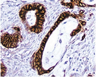
ICC Fixation Buffer is useful for fixing unstained cells that have been adhered to microscopic slides for cytokine immunocytochemical staining.  This fixation buffer is intended to preserve human and rodent lymphoid cells for the subsequent immunocytochemical staining of intracellular cytokines.  Once fixed the slides may be stored at -80°C for up to two months . When stored under those conditions no changes in cell morphology have been observed and the levels of intracellular cytokine detected by immunocytochemistry remain unaffected.

Note: The suitability of fixation conditions for cells undergoing immunocytochemistry staining depends on the availability of compatible antibodies that can specifically detect their cognate antigens under the same fixation conditions. With respect to intracellular cytokines and chemokines, BD Pharmingen offers a panel of purified anti-cytokine antibodies optimized for use in immunocytochemistry of cells fixed with formalin.


Recommended Assay Procedures

Procedure for using the Cytokine ICC Fixation buffer in Immunocytochemistry:

1.        Harvest cells and wash them twice in PBS using centrifugation (400 x g for 5 min) to remove residual protein.

2.        Adjust the cell concentration at 4 -5 x 10^6 cells/ml in PBS.

3.        Place 20 µl of the cell suspension in each well of the adhesion slides and let them adhere at room temperature (RT) for 20 min. Please note         that the slides should be washed in PBS at RT for 5 min before transferring the cells.

4.        Fix cells on slides using ICC Fixation Buffer (Cat No. 550010) for 15 min at RT.

5.        Wash slides 2X in PBS with 5 min incubations.

6.        Block slides with PBS supplemented with 1% (w/v) BSA (Sigma) for 30 min at RT or 10 min at 37°C.

7.        Wash slides 2X in PBS and proceed with staining or air dry them and store them at -80°C for future use.

8.        Incubate slides with 20 µl of 1% goat serum and PBS with 0.1% (w/v) saponin for 30 min at RT.

9.        Wash slides 2X with PBS with 5 min incubations.

10.        Block endogenous peroxidase activity with Endogenous Peroxidase Blocking Buffer (20 µl/well) for 10 min at RT.

11.        Wash 2X in PBS with 5 min incubations.

12.        Incubate each well with Avidin (20 µl/well) for 15 min.

13.        Wash 2X in PBS with 5 min incubations.

14.        Incubate each well with Biotin (20 µl/well) for 15 min.

15.        Wash 2X in PBS with 5 min incubations.

16.        Incubate each well for 1 hr at RT with 20 µl of purified cytokine-specific antibody or appropriate immunoglobulin isotype control diluted in         Antibody Diluent Buffer for IHC (Cat. No.559148).

17.        Wash slides 2X in PBS with 5 min incubations.

18.        Incubate each well with 20 µl of a biotinylated secondary antibody diluted in  Diluent Buffer (Cat. No. 559148) for 30 min at RT.

19.        Wash 2X in PBS with 5 min incubations.

20.        Apply 20 µl of Streptavidin-HRP (Cat. No. 550946). to each well on slides and incubate for 30 min at RT.

21.        Wash slides 2X with PBS with 5 minutes incubations.

22.        Incubate with 3-3´-Diaminobenzidine tetra hydrochloride (DAB), (Cat. No. 550880) for less than 5 min at RT.

23.        Stop the development of the color reaction by washing with PBS.

24.        The slides are subsequently mounted in short-term storage mounting medium.

Cytospins

1.        Assemble the Cytospin's sample chamber (e.g. Cytospin 3, Shandon, UK or comparable centrifuge), filter card, slide and cytospin racks         according to manufacturer's specifications.

2.        Load 40 µl of approximately 1 x 10^6 cells to each sample chamber.

3.        Spin slides at 600 rpm for 2 min.

4.        Take slides out of the cytospin rack and place them on a staining rack.

5.        For fixation and staining please follow the steps 4 through 24 specified above for staining cells on adhesion slides.

Danger:  ICC Fixation Buffer contains 2.09% formaldehyde.

Hazard statements

May cause an allergic skin reaction.

Suspected of causing genetic defects.

May cause cancer. Route of exposure: Inhalative.

Precautionary statements

Wear protective clothing / eye protection.

Wear protective gloves.

Avoid breathing mist/vapours/spray.

If skin irritation or rash occurs: Get medical advice/attention.

IF ON SKIN: Wash with plenty of water.

Dispose of contents/container in accordance with local/regional/national/international regulations.

Product Notices

  1. Please refer to www.bdbiosciences.com/us/s/resources for technical protocols.
550010 Rev. 2
Citations & References
Down Arrow Up Arrow

Development References (10)

  1. Andersson J, Abrams J, Bjork L, et al. Concomitant in vivo production of 19 different cytokines in human tonsils. Immunology. 1994; 83(1):16-24. (Biology). View Reference
  2. Andersson J, Bjork L, Dinarello CA, Towbin H, Andersson U. Lipopolysaccharide induces human interleukin-1 receptor antagonist and interleukin-1 production in the same cell. Eur J Immunol. 1992; 22(10):2617-2623. (Biology). View Reference
  3. Andersson J, Fehniger TE, Patterson BK, et al. Early reduction of immune activation in lymphoid tissue following highly active HIV therapy. AIDS. 1998; 12(11):F123-F129. (Biology). View Reference
  4. Barnes PF, Lu S, Abrams JS, Wang E, Yamamura M, Modlin RL. Cytokine production at the site of disease in human tuberculosis. Infect Immun. 1993; 61(8):3482-3489. (Biology). View Reference
  5. Bjork L, Fehniger TE, Andersson U, Andersson J. Computerized assessment of production of multiple human cytokines at the single-cell level using image analysis. J Leukoc Biol. 1996; 59(2):287-295. (Biology). View Reference
  6. Bjork L. Development of an image analysis system for the evaluation of cytokine production. Stockholm: 1995.
  7. Hsu SM, Raine L, Fanger H. A comparative study of the peroxidase-antiperoxidase method and an avidin-biotin complex method for studying polypeptide hormones with radioimmunoassay antibodies. Am J Clin Pathol. 1981; 75(5):734-738. (Biology). View Reference
  8. Hsu SM, Raine L, Fanger H. Use of avidin-biotin-peroxidase complex (ABC) in immunoperoxidase techniques: a comparison between ABC and unlabeled antibody (PAP) procedures. J Histochem Cytochem. 1981; 29(4):577-580. (Biology). View Reference
  9. Raqib R, Lindberg AA, Bjork L, et al. Down-regulation of gamma interferon, tumor necrosis factor type I, interleukin 1 (IL-1) type I, IL-3, IL-4, and transforming growth factor beta type I receptors at the local site during the acute phase of Shigella infection. Infect Immun. 1995; 63(8):3079-3087. (Biology). View Reference
  10. Sander B, Andersson J, Andersson U. Assessment of cytokines by immunofluorescence and the paraformaldehyde-saponin procedure. Immunol Rev. 1991; 119:65-93. (Biology). View Reference
View All (10) View Less
550010 Rev. 2

Please refer to Support Documents for Quality Certificates

Global - Refer to manufacturer's instructions for use and related User Manuals and Technical data sheets before using this products as described

Comparisons, where applicable, are made against older BD Technology, manual methods or are general performance claims.  Comparisons are not made against non-BD technologies, unless otherwise noted.

For Research Use Only. Not for use in diagnostic or therapeutic procedures.