Skip to main content Skip to navigation
CD33 (P67.6) APC-R700
Product Details
Down Arrow Up Arrow


BD®
Human
Mouse BALB/c IgG1, κ
CD33-transfected FMY9S5 Cells
5 µl
IV M503
CE_IVD


Preparation And Storage

Store undiluted at 4°C and protected from prolonged exposure to light. Do not freeze. The monoclonal antibody was purified from tissue culture supernatant or ascites by affinity chromatography. The antibody was conjugated to the dye under optimum conditions and unconjugated antibody and free dye were removed.

The antibody reagent is stable until the expiration date shown on the label when stored at 2° to 8°C. Do not use after the expiration date. Do not freeze the reagent or expose it to direct light during storage or incubation with cells. Keep the outside of the reagent vial dry.Do not use the reagent if you observe any change in appearance. Precipitation or discoloration indicates instability or deterioration.

Product Notices

  1. Please refer to www.bdbiosciences.com/us/s/resources for technical protocols.
  2. Alexa Fluor® is a registered trademark of Molecular Probes, Inc., Eugene, OR.
  3. Caution: Sodium azide yields highly toxic hydrazoic acid under acidic conditions. Dilute azide compounds in running water before discarding to avoid accumulation of potentially explosive deposits in plumbing.
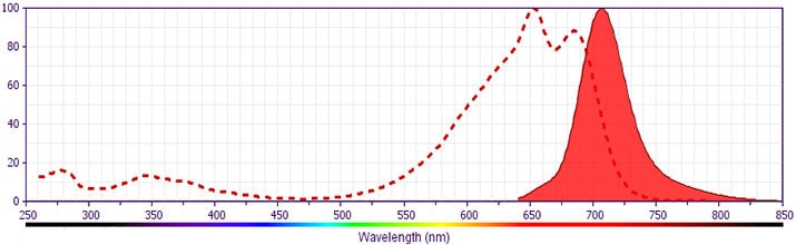
  4. For fluorochrome spectra and suitable instrument settings, please refer to our Multicolor Flow Cytometry web page at www.bdbiosciences.com/colors.
  5. Please refer to http://regdocs.bd.com to access safety data sheets (SDS).
  6. This reagent has been pre-diluted for use at the recommended Volume per Test. We typically use 1 × 10^6 cells in a 100-µl experimental sample (a test).
  7. Human donor specific background has been observed in relation to the presence of anti-polyethylene glycol (PEG) antibodies, developed as a result of certain vaccines containing PEG, including some COVID-19 vaccines. We recommend use of BD Horizon Brilliant™ Stain Buffer in your experiments to help mitigate potential background. For more information visit https://www.bdbiosciences.com/en-us/support/product-notices.
664455 Rev. 1
Antibody Details
Down Arrow Up Arrow
P67.6
664455 Rev. 1
Format Details
Down Arrow Up Arrow
APC-R700
The BD Horizon™ APC-R700 (APC-R700) Dye is a part of the BD APC red family of dyes. This tandem fluorochrome is comprised of an Allophycocyanin (APC) dye donor that has excitation maximum (Ex Max) of 651-nm and an acceptor dye, R700, with an emission maximum (Em Max) at 706-nm. APC-R700, driven by BD innovation, is designed to be excited by the red (627–640-nm) laser and detected using an optical filter centered near 710-nm (e.g., a 720/40-nm bandpass filter). APC-R700 is a brighter alternative to Alexa Fluor™ 700. Please ensure that your instrument’s configurations (lasers and optical filters) are appropriate for this dye.
altImg
APC-R700
Red 627-640 nm
651 nm
706 nm
664455 Rev.1
Citations & References
Down Arrow Up Arrow
View product citations for antibody "664455" on CiteAb

Development References (18)

  1. Andrews RG, Takahashi M, Segal GM, Powell JS, Bernstein ID, Singer JW. The L4F3 antigen is expressed by unipotent and multipotent colony-forming cells but not by their precursors. Blood. 1986; 68:1030-1035.
  2. Andrews RG, Torok-Storb B, Bernstein ID. Myeloid-associated differentiation antigens on stem cells and their progeny identified by monoclonal antibodies.. Blood. 1983; 62(1):124-32. View Reference
  3. Bernstein ID, Singer JW, Andrews RG, et al. Treatment of acute myeloid leukemia cells in vitro with a monoclonal antibody recognizing a myeloid differentiation antigen allows normal progenitor cells to be expressed.. J Clin Invest. 1987; 79(4):1153-9. View Reference
  4. Centers for Disease Control and Prevention. 2007 Guideline for Isolation Precautions: Preventing Transmission of Infectious Agents in Healthcare Settings. Available: https://www.cdc.gov/infectioncontrol/guidelines/isolation/index.html March 12, 2019. View Reference
  5. Churchill HRO, Fuda FS, Xu J, et al. Leukocyte immunoglobulin-like receptor B1 and B4 (LILRB1 and LILRB4): Highly sensitive and specific markers of acute myeloid leukemia with monocytic differentiation.. Cytometry B Clin Cytom. 2021; 100(4):476-487. View Reference
  6. Clinical and Laboratory Standards Institute. Clinical Flow Cytometric Analysis of Neoplastic Hematolymphoid Cells. In: CLSI. CLSI, ed. CLSI document H43-A2. Wayne, PA: Clinical and Laboratory Standards Institute; 2007:1-81. View Reference
  7. Clinical and Laboratory Standards Institute. Collection of Diagnostic Venous Blood Specimens, 7th ed. In: CLSI. CLSI, ed. CLSI document GP41-A7. Wayne, PA: Clinical and Laboratory Standards Institute; 2017:1-85. View Reference
  8. Clinical and Laboratory Standards Institute. Protection of Laboratory Workers from Occupationally Acquired Infections. In: CLSI. CLSI, ed. CLSI document M29-A4. Wayne, PA: Clinical and Laboratory Standards Institute; 2014:1-133. View Reference
  9. Dunphy CH, Tang W. The value of CD64 expression in distinguishing acute myeloid leukemia with monocytic differentiation from other subtypes of acute myeloid leukemia: a flow cytometric analysis of 64 cases.. Arch Pathol Lab Med. 2007; 131(5):748-54. View Reference
  10. Ehninger A, Kramer M, Röllig C, et al. Distribution and levels of cell surface expression of CD33 and CD123 in acute myeloid leukemia.. Blood Cancer J. 2014; 4(6):e218. View Reference
  11. Haycocks NG, Lawrence L, Cain JW, Zhao XF. Optimizing antibody panels for efficient and cost-effective flow cytometric diagnosis of acute leukemia.. Cytometry B Clin Cytom. 2011; 80(4):221-9. View Reference
  12. Jackson AL, Warner NL. Rose NR, Friedman H, Fahey JL, ed. Manual of Clinical Laboratory Immunology 3rd ed. Washington, DC: American Society for Microbiology; 1986:226-235.
  13. Kroll MH. Evaluating interference caused by lipemia.. Clin Chem. 2004; 50(11):1968-9. View Reference
  14. Köller U, Peschel CH. Cluster report: CD33. In: Knapp W. W. Knapp .. et al., ed. Leucocyte typing IV : white cell differentiation antigens. Oxford New York: Oxford University Press; 1989:812-813.
  15. Nikolac N. Lipemia: causes, interference mechanisms, detection and management.. Biochem Med (Zagreb). 2014; 24(1):57-67. View Reference
  16. Schwinzer R. Cluster Report: CD45/CD45R. In: Knapp W. W. Knapp .. et al., ed. Leucocyte typing IV : white cell differentiation antigens. Oxford New York: Oxford University Press; 1989:628-634.
  17. Stelzer GT, Marti G, Hurley A, McCoy P, Lovett EJ, Schwartz A. U.S.-Canadian Consensus recommendations on the immunophenotypic analysis of hematologic neoplasia by flow cytometry: standardization and validation of laboratory procedures.. Cytometry. 1997; 30(5):214-30. View Reference
  18. Terstappen LW, Hollander Z, Meiners H, Loken MR. Quantitative comparison of myeloid antigens on five lineages of mature peripheral blood cells. J Leukoc Biol. 1990; 48(2):138-148. View Reference
View All (18) View Less
664455 Rev. 1

Please refer to Support Documents for Quality Certificates


Global - Refer to manufacturer's instructions for use and related User Manuals and Technical data sheets before using this products as described


Comparisons, where applicable, are made against older BD Technology, manual methods or are general performance claims.  Comparisons are not made against non-BD technologies, unless otherwise noted.

For Research Use Only. Not for use in diagnostic or therapeutic procedures.

Documents are subject to revision without notice. Please verify you have the correct revision of the document, and always refer back to BD's eIFU website for the latest and most up to date information.