

Preparation And Storage
Recommended Assay Procedures
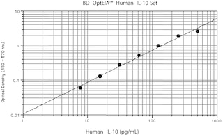
1. ELISA Capture: The purified JES3-19F1 antibody (Cat. No. 554705) is useful as a capture antibody for a sandwich ELISA for specifically measuring human IL-10 protein levels. Purified JES3-19F1 antibody (Cat. No. 554705) can be paired with the biotinylated JES3-12G8 antibody (Cat. No. 554499) as the detection antibody and with recombinant human IL-10 protein (Cat. No. 554611) as the standard. The purified JES3-19F1 capture antibody should be titrated 1-4 µg/ml to determine optimal concentration for ELISA capture. To obtain linear standard curves, doubling dilutions of human IL-10 ranging from ~2,000 to 15 pg/ml are recommended for inclusion in each ELISA plate. For specific methodology, please visit the protocols section or chapter on ELISA in the Immune Function Handbook, both of which are posted on our web site, www.bdbiosciences.com.
Note 1: This ELISA pair shows no cross-reactivity with any of the cytokines tested ( e.g., mouse IL-1ß, IL-2, IL-3, IL-4, IL-5, IL-6, IL-7, IL-9, IL-10, IL-12 p70, IL-15, GM-CSF, IFN-γ, MCP-1, TCA-3, TNF; human IL-1α, IL-1 ß, IL-2, IL-3, IL-4, IL-5, IL-6, IL-7, IL-8, IL-9, IL-11, IL-12 p70, IL-12 p40, IL-13, IL-15, G-CSF, GM-CSF, IFN-γ, lymphotactin, MCP-1, MCP-2, MIP-1α, MIP-1ß, NT-3, PDGF-AA, sCD23 , SCF, TNF, LT-α, VEGF; rat IL-2, IL-4, IL-6, IL-10, GM-CSF, IFN-γ, TNF).
Note 2: This ELISA pair is recommended primarily for measuring cytokine from experimental cell culture systems. These ELISA reagents are not recommended for assaying serum samples For testing human IL-10 in complex biological fluids like serum or plasma, our human IL-10 OptEIA™ ELISA Set (Cat. No. 555157) and OptEIA™ ELISA Kit (Cat. No. 550613) are recommended.
2. IF/Flow: The JES3-19F1 antibody is useful for immunofluorescent staining and flow cytometric analysis to identify and enumerate IL-10 producing cells within mixed cell populations. The directly-conjugated JES3-19F1 antibodies are especially suitable for these studies. For specific methodology, please visit the protocols section or chapter on intracellular staining in the Immune Function Handbook, both of which are posted on our web site, www.bdbiosciences.com.
3. Neutralization: The JES3-19F1 antibody is useful for neutralization of human IL-10 bioactivity. The purified JES319F1 antibody is supplied in our NA/LE™ format (Cat. No. 554703), which is sodium azide-free, sterile-filtered (0.2 µm pore), sterility-tested, and with assured low endotoxin for this purpose. Endotoxin level as determined by LAL assay is less than 0.01 ng/µg protein. A suitable NA/LE rat IgG2a isotype control to match the NA/LE JES3-19F1 antibody is the R35-95 antibody, (Cat. No. 554687).
Product Notices
- Since applications vary, each investigator should titrate the reagent to obtain optimal results.
- An isotype control should be used at the same concentration as the antibody of interest.
- Caution: Sodium azide yields highly toxic hydrazoic acid under acidic conditions. Dilute azide compounds in running water before discarding to avoid accumulation of potentially explosive deposits in plumbing.
- Species cross-reactivity detected in product development may not have been confirmed on every format and/or application.
- Please refer to http://regdocs.bd.com to access safety data sheets (SDS).
- Sodium azide is a reversible inhibitor of oxidative metabolism; therefore, antibody preparations containing this preservative agent must not be used in cell cultures nor injected into animals. Sodium azide may be removed by washing stained cells or plate-bound antibody or dialyzing soluble antibody in sodium azide-free buffer. Since endotoxin may also affect the results of functional studies, we recommend the NA/LE (No Azide/Low Endotoxin) antibody format, if available, for in vitro and in vivo use.
- Please refer to www.bdbiosciences.com/us/s/resources for technical protocols.
Companion Products


The JES3-19F1 monoclonal antibody specifically recognizes human Interleukin-10 (IL-10) that is encoded by IL10. IL-10 is also known as Cytokine Synthesis Inhibitory Factor (CSIF), B cell-derived T cell growth factor (B-TCGF), and T-cell growth inhibitory factor (TGIF). The JES3-19F1 antibody crossreacts with ebvIL-10 protein, the Epstein-Barr viral IL-10 homolog (viral IL-10 or vIL-10) encoded by the BCRF1 gene. IL-10 is produced by a variety of cells such as some activated T cells and B cells including regulatory T cells (Treg) and B cells (Breg), monocytes and macrophages, dendritic cells (DC), keratinocytes, and mast cells. IL-10 is a multifunctional cytokine that can downregulate immune and proinflammatory responses. IL-10 can act to reduce expression of major histocompatibility complex class II antigens, costimulatory molecules, or proinflammatory cytokines including IL-1β, IL-2, IL-3, IL-12, IFN-γ, TNF or GM-CSF expressed by activated monocytes, macrophages, dendritic cells (DC), natural killer (NK) cells, or T cells. IL-10 has been shown to play a role in chronic viral infections. IL-10 can also enhance B cell survival, proliferation, and differentiation to become antibody-producing cells. The JES3-19F1 antibody reportedly neutralizes the biological activity of human IL-10 and ebvIL-10. IL-10 mediates its biological activities by signaling through a heterotetrameric receptor complex composed of the type II cytokine receptor subunits CD210a (IL-10 Rα) and CD210b (IL-10 Rβ).
Development References (3)
-
Andersson EC, Christensen JP, Marker O, Thomsen AR. Changes in cell adhesion molecule expression on T cells associated with systemic virus infection. Immunology. 1994; 152(3):1237-1245. (Clone-specific). View Reference
-
D'Andrea A, Aste-Amezaga M, Valiante NM, Ma X, Kubin M, Trinchieri G. Interleukin 10 (IL-10) inhibits human lymphocyte interferon gamma-production by suppressing natural killer cell stimulatory factor/IL-12 synthesis in accessory cells. J Exp Med. 1993; 178(3):1041-1048. (Clone-specific: Neutralization). View Reference
-
Prussin C, Metcalfe DD. Detection of intracytoplasmic cytokine using flow cytometry and directly conjugated anti-cytokine antibodies. J Immunol Methods. 1995; 188(1):117-128. (Methodology). View Reference
Please refer to Support Documents for Quality Certificates
Global - Refer to manufacturer's instructions for use and related User Manuals and Technical data sheets before using this products as described
Comparisons, where applicable, are made against older BD Technology, manual methods or are general performance claims. Comparisons are not made against non-BD technologies, unless otherwise noted.
For Research Use Only. Not for use in diagnostic or therapeutic procedures.