Old Browser
This page has been recently translated and is available in French now.
Looks like you're visiting us from {countryName}.
Would you like to stay on the current country site or be switched to your country?


Anti-human p53, Cat. No. 554294. Formalin-fixed, paraffin-embedded tissue section of human breast carcinoma stained with anti-human p53 (clone DO-7) using a DAB chromogen and Hematoxylin counterstain.

Western blot analysis of p53 in CEM human leukemia cell lysates. Lane 1, clone DO-7 (Cat. No. 554294). Lane 2, a mouse IgG2b isotype control.

Profile of permeabilized CEM human leukemia cells analyzed on a FACScan™ (BDIS, San Jose, CA). Cells were stained with anti-human p53 (clone DO-7) or a mouse IgG2b isotype control followed by FITC-conjugated second step.


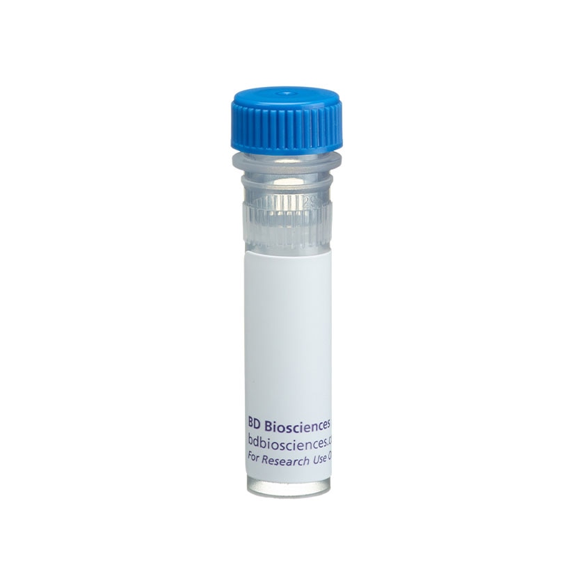
BD Pharmingen™ Purified Mouse Anti-Human p53

BD Pharmingen™ Purified Mouse Anti-Human p53

BD Pharmingen™ Purified Mouse Anti-Human p53

Regulatory Status Legend
Any use of products other than the permitted use without the express written authorization of Becton, Dickinson and Company is strictly prohibited.
Preparation And Storage
Recommended Assay Procedures
Positive control cell lines include SK-BR-3 breast carcinoma cells (ATCC HTB-30), A431 epidermal carcinoma cells (ATCC CRL-1555) and CEM human leukemia cells (ATCC CCL-119). MCF-7 breast carcinoma cells (ATCC HTB-22) are suggested as a negative control. Positive immunostaining is seen in a high proportion of breast and colon carcinomas. p53 staining is not typically detected in normal skin, brain, kidney, lung, stomach, or breast tissue.
Product Notices
- Since applications vary, each investigator should titrate the reagent to obtain optimal results.
- Caution: Sodium azide yields highly toxic hydrazoic acid under acidic conditions. Dilute azide compounds in running water before discarding to avoid accumulation of potentially explosive deposits in plumbing.
- Sodium azide is a reversible inhibitor of oxidative metabolism; therefore, antibody preparations containing this preservative agent must not be used in cell cultures nor injected into animals. Sodium azide may be removed by washing stained cells or plate-bound antibody or dialyzing soluble antibody in sodium azide-free buffer. Since endotoxin may also affect the results of functional studies, we recommend the NA/LE (No Azide/Low Endotoxin) antibody format, if available, for in vitro and in vivo use.
- Species cross-reactivity detected in product development may not have been confirmed on every format and/or application.
- Please refer to www.bdbiosciences.com/us/s/resources for technical protocols.
p53 is a 53 kD nuclear phosphoprotein that acts as a tumor suppressor protein, and is involved in inhibiting cell proliferation when DNA damage occurs. The gene for p53 is the most commonly mutated gene yet identified in human cancers. Missense mutations occur in tumors of the colon, lung, breast, ovary, bladder and several other organs. The mutant p53 is overexpressed in a variety of transformed cells and wildtype p53 forms specific complexes with several viral oncogenes including SV40 large T, E1B from adenovirus, and E6 from human papilloma virus. Wildtype p53 plays a role as a checkpoint protein for DNA damage during the G1/S-phase of the cell cycle. However, it is still unclear, whether point mutated forms of p53 are simple null mutants and/or dominant negatively acting proteins.
The DO-7 antibody recognizes human wildtype and mutant p53. It cross-reacts with bovine p53 but does not cross-react with mouse or rat p53. DO-7 recognizes an epitope between amino acids 1-45 of known forms of human p53. Human recombinant p53 protein was used as immunogen. Wildtype 53 proteins have a very short half-life and are usually not detectable with monoclonal antibodies (mAbs) in normal tissues.
Development References (7)
-
Baas IO, Mulder JW, Offerhaus GJ, Vogelstein B, Hamilton SR. An evaluation of six antibodies for immunohistochemistry of mutant p53 gene product in archival colorectal neoplasms. J Pathol. 1994; 172(1):5-12. (Clone-specific: Immunohistochemistry). View Reference
-
Bonsing BA, Corver WE, Gorsira MC, et al. Specificity of seven monoclonal antibodies against p53 evaluated with Western blotting, immunohistochemistry, confocal laser scanning microscopy, and flow cytometry. Cytometry. 1997; 28(1):11-24. (Clone-specific: Flow cytometry). View Reference
-
Cripps KJ, Purdie CA, Carder PJ, et al. A study of stabilisation of p53 protein versus point mutation in colorectal carcinoma. Oncogene. 1994; 9(9):2739-2743. (Clone-specific: Immunohistochemistry). View Reference
-
Jacquemier J, Moles JP, Penault-Llorca F, et al. p53 immunohistochemical analysis in breast cancer with four monoclonal antibodies: comparison of staining and PCR-SSCP results. Br J Cancer. 1994; 69(5):846-852. (Clone-specific: Immunohistochemistry). View Reference
-
Lambkin HA, Mothersill CM, Kelehan P. Variations in immunohistochemical detection of p53 protein overexpression in cervical carcinomas with different antibodies and methods of detection. J Pathol. 1994; 172(1):13-18. (Clone-specific: Immunohistochemistry). View Reference
-
Vogelstein B. Cancer. A deadly inheritance. Nature. 1990; 348(6303):681-682. (Biology). View Reference
-
Vojtesek B, Bartek J, Midgley CA, Lane DP. An immunochemical analysis of the human nuclear phosphoprotein p53. New monoclonal antibodies and epitope mapping using recombinant p53. J Immunol Methods. 1992; 151(1-2):237-244. (Clone-specific: Immunohistochemistry, Immunoprecipitation, Western blot). View Reference
Please refer to Support Documents for Quality Certificates
Global - Refer to manufacturer's instructions for use and related User Manuals and Technical data sheets before using this products as described
Comparisons, where applicable, are made against older BD Technology, manual methods or are general performance claims. Comparisons are not made against non-BD technologies, unless otherwise noted.
For Research Use Only. Not for use in diagnostic or therapeutic procedures.
Report a Site Issue
This form is intended to help us improve our website experience. For other support, please visit our Contact Us page.