





.png)


Preparation And Storage


Product Notices
- Since applications vary, each investigator should titrate the reagent to obtain optimal results.
- Please refer to www.bdbiosciences.com/us/s/resources for technical protocols.
- For fluorochrome spectra and suitable instrument settings, please refer to our Multicolor Flow Cytometry web page at www.bdbiosciences.com/colors.
- Caution: Sodium azide yields highly toxic hydrazoic acid under acidic conditions. Dilute azide compounds in running water before discarding to avoid accumulation of potentially explosive deposits in plumbing.
Companion Products
.png?imwidth=320)
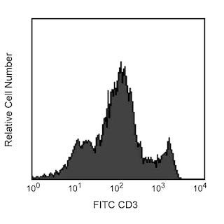
The OX-8 monoclonal antibody specifically binds to the hinge-like membrane-proximal domain of the 32 kDa α chain of the CD8 differentiation antigen. A truncated CD8 α' isoform has not been detected in the rat. The CD8 α and β chains (CD8a and CD8b, respectively) form a heterodimer on the surface of most thymocytes and a subpopulation of mature T lymphocytes (i.e., MHC class I-restricted T cells, including most T suppressor/cytotoxic cells). Intestinal intrapithelial lymphocytes, many CD8+ T cells of athymic rats, many activated CD4+ T cells, and most NK cells express CD8a without CD8b. It has been suggested that the expression of the CD8a/CD8b heterodimer is restricted to thymus-derived T lymphocytes. OX-8 antibody does not react with resting CD4+ T helper cells. CD8 is an antigen coreceptor on the T-cell surface which interacts with MHC class I molecules on antigen-presenting cells. It participates in T-cell activation through its association with the T-cell receptor complex and protein tyrosine kinase Ick. Macrophages have also been reported to express CD8 α and β chains, which are involved in signal transduction. Soluble OX-8 mAb partially blocks in vitro MLR and CTL activity.

Development References (15)
-
Barclay AN. The localization of populations of lymphocytes defined by monoclonal antibodies in rat lymphoid tissues. J Immunol. 1981; 42(4):593-600. (Clone-specific: Immunohistochemistry). View Reference
-
Bierer BE, Sleckman BP, Ratnofsky SE, Burakoff SJ. The biologic roles of CD2, CD4, and CD8 in T-cell activation. Annu Rev Immunol. 1989; 7:579-599. (Biology). View Reference
-
Brideau RJ, Carter PB, McMaster WR, Mason DW, Williams AF. Two subsets of rat T lymphocytes defined with monoclonal antibodies.. Eur J Immunol. 1980; 10:609-615. (Immunogen: Flow cytometry). View Reference
-
Classon BJ, Brown MH, Garnett D, et al. The hinge region of the CD8 alpha chain: structure, antigenicity, and utility in expression of immunoglobulin superfamily domains. Int Immunol. 1992; 4(2):215-225. (Clone-specific). View Reference
-
Hirji N, Lin TJ, Befus AD. A novel CD8 molecule expressed by alveolar and peritoneal macrophages stimulates nitric oxide production. J Immunol. 1997; 158(4):1833-1840. (Clone-specific: Stimulation). View Reference
-
Hirji N, Lin TJ, Bissonnette E, Belosevic M, Befus AD. Mechanisms of macrophage stimulation through CD8: macrophage CD8alpha and CD8beta induce nitric oxide production and associated killing of the parasite Leishmania major. J Immunol. 1998; 160(12):6004-6011. (Clone-specific: Stimulation). View Reference
-
Janeway CA Jr. The T cell receptor as a multicomponent signalling machine: CD4/CD8 coreceptors and CD45 in T cell activation. Annu Rev Immunol. 1992; 10:645-674. (Biology). View Reference
-
Johnson P, Gagnon J, Barclay AN, Williams AF. Purification, chain separation and sequence of the MRC OX-8 antigen, a marker of rat cytotoxic T lymphocytes. EMBO J. 1985; 4(10):2539-2545. (Clone-specific: Immunoaffinity chromatography). View Reference
-
Kuhnlein P, Park JH, Herrmann T, Elbe A, Hunig T. Identification and characterization of rat gamma/delta T lymphocytes in peripheral lymphoid organs, small intestine, and skin with a monoclonal antibody to a constant determinant of the gamma/delta T cell receptor. J Immunol. 1994; 153(3):979-986. (Biology). View Reference
-
Mason DW, Arthur RP, Dallman MJ, Green JR, Spickett GP, Thomas ML. Functions of rat T-lymphocyte subsets isolated by means of monoclonal antibodies. Immunol Rev. 1983; 74:57-82. (Clone-specific: Blocking). View Reference
-
Mitnacht R, Bischof A, Torres-Nagel N, Hunig T. Opposite CD4/CD8 lineage decisions of CD4+8+ mouse and rat thymocytes to equivalent triggering signals: correlation with thymic expression of a truncated CD8 alpha chain in mice but not rats. J Immunol. 1998; 160(2):700-707. (Clone-specific: Immunoprecipitation, Western blot). View Reference
-
Scriba A, Grau V, Steiniger B. Phenotype of rat monocytes during acute kidney allograft rejection: increased expression of NKR-P1 and reduction of CD43. Scand J Immunol. 1998; 47(4):332-342. (Biology). View Reference
-
Thomas ML, Green JR. Molecular nature of the W3/25 and MRC OX-8 marker antigens for rat T lymphocytes: comparisons with mouse and human antigens. Eur J Immunol. 1983; 13(10):855-858. (Clone-specific: Immunoprecipitation). View Reference
-
Torres-Nagel N, Kraus E, Brown MH, et al. Differential thymus dependence of rat CD8 isoform expression.. Eur J Immunol. 1992; 22(11):2841-2848. (Clone-specific: Blocking, Immunoprecipitation, Western blot). View Reference
-
Wallgren AC, Karlsson-Parra A, Korsgren O. The main infiltrating cell in xenograft rejection is a CD4+ macrophage and not a T lymphocyte. Transplantation. 1995; 60(6):594-601. (Clone-specific: Immunohistochemistry). View Reference
Please refer to Support Documents for Quality Certificates
Global - Refer to manufacturer's instructions for use and related User Manuals and Technical data sheets before using this products as described
Comparisons, where applicable, are made against older BD Technology, manual methods or are general performance claims. Comparisons are not made against non-BD technologies, unless otherwise noted.
For Research Use Only. Not for use in diagnostic or therapeutic procedures.
