Skip to main content Skip to navigation
PE Mouse Anti-Human MIP-1α (CCL3)
Flow cytometric analysis of MIP-1α (CCL3) expression by stimulated CD14+ human monocytes. Human PBMC were stimulated for 6 hr with LPS (10 ng/ml) in the presence of GolgiStop™ (Cat. No. 554724; aka monensin 2 µM). The PBMC were harvested, stained with FITC Mouse Anti-Human CD14 (Cat. No. 555397), fixed, permeabilized, and subsequently stained with 0.25 µg of PE Mouse Anti-Human MIP-1α (CCL3) (Cat. No. 554730) following Pharmingen's staining protocol (left panel). The data reflect gating on monocytes, based on forward and side light scatter. To demonstrate specificity of staining, the binding of PE Mouse Anti-Human MIP-1α (CCL3) was blocked by the preincubation of the conjugated antibody with recombinant human MIP-1α (0.5 µg; middle panel), and by preincubation of the fixed/permeabilized cells with the unlabeled 11A3 antibody (5.0 µg, right panel) prior to staining with the conjugate antibody. The quadrant markers for the bivariate dot plots were set based on the autofluorescence control, and verified with the recombinant cytokine blocking (middle panel) and unlabeled antibody (right panel) blocking specificity controls.
Product Details
Down Arrow Up Arrow


BD Pharmingen™
CCL3
Human (QC Testing), Rhesus, Cynomolgus (Tested in Development)
Mouse IgG2a, κ
Intracellular staining (flow cytometry) (Routinely Tested), Immunofluorescence (Tested During Development)
0.2 mg/ml
AB_395532
Aqueous buffered solution containing ≤0.09% sodium azide.
RUO


Preparation And Storage

Store undiluted at 4°C and protected from prolonged exposure to light. Do not freeze. The monoclonal antibody was purified from tissue culture supernatant or ascites by affinity chromatography. The antibody was conjugated with R-PE under optimum conditions, and unconjugated antibody and free PE were removed.

Recommended Assay Procedures

The PE Mouse Anti-Human MIP-1α (CCL3) can be used for multicolor immunofluorescent staining and flow cytometric analyses to identify and enumerate MIP-1α (CCL3) producing cells with mixed cell populations. For optimal immunofluorescent staining with flow cytometric analysis, this anti-cytokine antibody should be titrated (≤ 0.5 µg mAb/million cells).

A useful control for demonstrating specificity of staining is either of the following: 1) pre-block the conjugated 11A3 antibody with ligand (e.g., recombinant human MIP-1α (CCL3)) prior to staining, or 2) pre-block the fixed/permeabilized cells with unlabeled 11A3 antibody prior to staining. The intracellular staining technique and use of blocknig controls are described in detail by C. Prussin and D. Metcalfe. A suitable mouse IgG2a, κ isotype control immunoglobulin for assessing the level of background staining on paraformaldehyde-fixed/saponin-permabilized human cells is PE Mouse IgG2a, κ Isotype Control (Cat. No. 554648); use at comparable concentrations to antibody of interest (e.g., ≤0.5 µg mAb/1 million cells).

Product Notices

  1. Since applications vary, each investigator should titrate the reagent to obtain optimal results.
  2. An isotype control should be used at the same concentration as the antibody of interest.
  3. Caution: Sodium azide yields highly toxic hydrazoic acid under acidic conditions. Dilute azide compounds in running water before discarding to avoid accumulation of potentially explosive deposits in plumbing.
  4. For fluorochrome spectra and suitable instrument settings, please refer to our Multicolor Flow Cytometry web page at www.bdbiosciences.com/colors.
  5. Species cross-reactivity detected in product development may not have been confirmed on every format and/or application.
  6. Please refer to http://regdocs.bd.com to access safety data sheets (SDS).
  7. Please refer to www.bdbiosciences.com/us/s/resources for technical protocols.
554730 Rev. 3
Antibody Details
Down Arrow Up Arrow
11A3

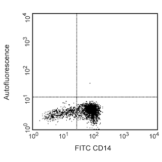
The 11A3 monoclonal antibody specifically recognizes macrophage inflammatory protein-1α (MIP-1α), a member of the CC chemokine family of proteins. It is produced by T cells, B cells, Langerhans cells, neutrophils and macrophages. MIP-1α plays a role as an inhibitor of stem cell proliferation and as a chempattractant of B cells, eosinophils and cytotoxic T cells. Clone 11A3 also cross-reacts with an intracellular component of rhesus and cynomolgus macaque-LPS-stimulated peripheral blood monocytes. The reactivity pattern observed on CD14-positive cells is similar to that seen on normal human peripheral blood monocytes.

554730 Rev. 3
Format Details
Down Arrow Up Arrow
PE
R-Phycoerythrin (PE), is part of the BD family of Phycobiliprotein dyes. This fluorochrome is a multimeric fluorescent phycobiliprotein with excitation maximum (Ex Max) of 496 nm and 566 nm and an emission maximum (Em Max) at 576 nm. PE is designed to be excited by the Blue (488 nm), Green (532 nm) and Yellow-Green (561 nm) lasers and detected using an optical filter centered near 575 nm (e.g., a 575/26-nm bandpass filter). As PE is excited by multiple lasers, this can result in cross-laser excitation and fluorescence spillover on instruments with various combinations of Blue, Green, and Yellow-Green lasers. Please ensure that your instrument’s configurations (lasers and optical filters) are appropriate for this dye.
altImg
PE
Yellow-Green 488 nm, 532 nm, 561 nm
496 nm, 566 nm
576 nm
554730 Rev.3
Citations & References
Down Arrow Up Arrow

Development References (4)

  1. Prussin C, Metcalfe DD. Detection of intracytoplasmic cytokine using flow cytometry and directly conjugated anti-cytokine antibodies. J Immunol Methods. 1995; 188(1):117-128. (Biology: Flow cytometry). View Reference
  2. Rollins BJ. Chemokines. Blood. 1997; 90(3):909-928. (Biology). View Reference
  3. Vaddi K, Keller M, Newton RC. The chemokine factsbook. San Diego: Academic Press; 1997:205 p.
  4. Wolpe SD, Cerami A. Macrophage inflammatory proteins 1 and 2: members of a novel superfamily of cytokines. FASEB J. 1989; 3(14):2565-2573. (Biology). View Reference
View All (4) View Less
554730 Rev. 3

Please refer to Support Documents for Quality Certificates


Global - Refer to manufacturer's instructions for use and related User Manuals and Technical data sheets before using this products as described


Comparisons, where applicable, are made against older BD Technology, manual methods or are general performance claims.  Comparisons are not made against non-BD technologies, unless otherwise noted.

For Research Use Only. Not for use in diagnostic or therapeutic procedures.