





.png)


Preparation And Storage


Recommended Assay Procedures
For more information about BD Phosflow™: Please refer to http://www.bdbiosciences.com/support/resources/phosflow/index.jsp
Investigators may also find the following protocols to be helpful:
Phosflow protocol for human PBMC: Please refer to http://www.bdbiosciences.com/documents/Phosflow_Protocol_for_Human_PBMCs.pdf
Phosflow protocol for human whole blood: Please refer to http://www.bdbiosciences.com/documents/Phosflow_Protocol_for_Human_Whole_Blood_Samples.pdf
Phosflow protocol for adherent cells: Please refer to http://www.bdbiosciences.com/support/resources/protocols/protocol_adherent.jsp
Product Notices
- This reagent has been pre-diluted for use at the recommended Volume per Test. We typically use 1 × 10^6 cells in a 100-µl experimental sample (a test).
- Please refer to www.bdbiosciences.com/us/s/resources for technical protocols.
- Caution: Sodium azide yields highly toxic hydrazoic acid under acidic conditions. Dilute azide compounds in running water before discarding to avoid accumulation of potentially explosive deposits in plumbing.
- Source of all serum proteins is from USDA inspected abattoirs located in the United States.
- For fluorochrome spectra and suitable instrument settings, please refer to our Multicolor Flow Cytometry web page at www.bdbiosciences.com/colors.
Companion Products

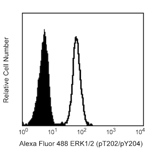


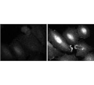
The members of the Mitogen-Activated Protein Kinase (MAPK) family are components of a key signal transduction cascade that links events at the cell surface to responses in the nucleus. The signaling cascade is found in species as varied as yeast and humans, with many of the proteins being well conserved. In mammals the most widely studied members of the cascade are the Extracellular signal-Regulated Kinases, ERK1 (p44 MAPK) and ERK2 (p42 MAPK). ERK1 and ERK2 share 85% homology and are activated by extracellular signals such as growth factors, hormones, and phorbol esters. Activation occurs through a series of phosphorylations by kinases activating other kinases and eventually leading to phosphorylation of the ERKs. Growth factor stimulation leads to activation of Ras and Raf, leading to phosphorylation of MEK1 (MAPK/ERK kinase) which, in turn, activates the ERKs via dual phosphorylation. Once activated, the ERKs phosphorylate other cytoplasmic signalling molecules, cell-surface receptors, microtubule-associated proteins, and transcription factors in the nucleus. Thus, the active ERK has myriad downstream effectors that implicate it in the control of cell proliferation and differentiation, as well as regulation of the cytoskeleton. Furthermore, studies have shown that elevated ERK activity is associated with some cancers.
The 20A monoclonal antibody recognizes the phosphorylated threonine 202 and tyrosine 204 (pT202/pY204) of human ERK1 and pT184/pY186 of human ERK2. The orthologous phosphorylation sites in murine ERK1 and ERK2 are T203/Y205 and T183/Y185.

Development References (3)
-
Boulton TG, Cobb MH. Identification of multiple extracellular signal-regulated kinases (ERKs) with antipeptide antibodies. Cell Regul. 1991; 2(5):357-371. (Biology). View Reference
-
Clark EA, Hynes RO. Ras activation is necessary for integrin-mediated activation of extracellular signal-regulated kinase 2 and cytosolic phospholipase A2 but not for cytoskeletal organization. J Biol Chem. 1996; 271(25):14814-14818. (Biology). View Reference
-
Sivaraman VS, Wang H, Nuovo GJ, Malbon CC. Hyperexpression of mitogen-activated protein kinase in human breast cancer. J Clin Invest. 1997; 99(7):1478-1483. (Biology). View Reference
Please refer to Support Documents for Quality Certificates
Global - Refer to manufacturer's instructions for use and related User Manuals and Technical data sheets before using this products as described
Comparisons, where applicable, are made against older BD Technology, manual methods or are general performance claims. Comparisons are not made against non-BD technologies, unless otherwise noted.
For Research Use Only. Not for use in diagnostic or therapeutic procedures.
