





.png)


Preparation And Storage


Product Notices
- Since applications vary, each investigator should titrate the reagent to obtain optimal results.
- Please refer to www.bdbiosciences.com/us/s/resources for technical protocols.
- For fluorochrome spectra and suitable instrument settings, please refer to our Multicolor Flow Cytometry web page at www.bdbiosciences.com/colors.
- Although hamster immunoglobulin isotypes have not been well defined, BD Biosciences Pharmingen has grouped Armenian and Syrian hamster IgG monoclonal antibodies according to their reactivity with a panel of mouse anti-hamster IgG mAbs. A table of the hamster IgG groups, Reactivity of Mouse Anti-Hamster Ig mAbs, may be viewed at http://www.bdbiosciences.com/documents/hamster_chart_11x17.pdf.
- Caution: Sodium azide yields highly toxic hydrazoic acid under acidic conditions. Dilute azide compounds in running water before discarding to avoid accumulation of potentially explosive deposits in plumbing.
Companion Products
.png?imwidth=320)
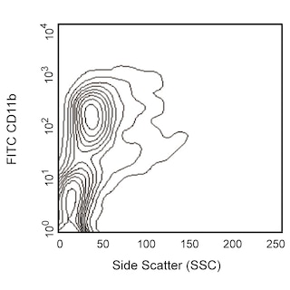
The HMα2 antibody reacts with integrin α2 chain (CD49b), the 150-kDa transmembrane glycoprotein that non-covalently associates with the integrin β1 subunit (CD29) to form the integrin α2β1 complex known as VLA-2. VLA-2, a receptor for collagen and laminin, is expressed on some splenic CD4+ T lymphocytes and NK-T cells, intestinal intraepithelial and lamina propria lymphocytes, splenic NK cells, epithelial cells, and platelets; but it is not on thymocytes or Peyer's-patch or lymphnode lymphocytes. The expression of VLA-2 is upregulated on lymphocytes in response to mitogens. The HMα2 antibody has been reported to partially block the interaction of T-cell blasts, but not NK cells, with collagen. Purified HMα2 mAb blocks the staining of splenic NK cells by the anti-CD49b/Pan-NK Cells mAb DX5 (Cat. No. 553858, for the PE conjugate). Therefore, mAb HMα2 may be used like the DX5 mAb for identification of NK cells.

Development References (5)
-
Arase H, Saito T, Phillips JH, Lanier LL. Cutting edge: the mouse NK cell-associated antigen recognized by DX5 monoclonal antibody is CD49b (alpha 2 integrin, very late antigen-2). J Immunol. 2001; 167(3):1141-1144. (Biology). View Reference
-
Chen H, Paul WE. A population of CD62Llow Nk1.1- CD4+ T cells that resembles NK1.1+ CD4+ T cells. Eur J Immunol. 1998; 28(10):3172-3182. (Biology). View Reference
-
Miyake S, Sakurai T, Okumura K, Yagita H. Identification of collagen and laminin receptor integrins on murine T lymphocytes. Eur J Immunol. 1994; 24(9):2000-2005. (Immunogen). View Reference
-
Noto K, Kato K, Okumura K, Yagita H. Identification and functional characterization of mouse CD29 with a mAb. Int Immunol. 1995; 7(5):835-842. (Biology). View Reference
-
Tanaka T, Ohtsuka Y, Yagita H, Shiratori Y, Omata M, Okumura K. Involvement of alpha 1 and alpha 4 integrins in gut mucosal injury of graft-versus-host disease. Int Immunol. 1995; 7(8):1183-1189. (Biology). View Reference
Please refer to Support Documents for Quality Certificates
Global - Refer to manufacturer's instructions for use and related User Manuals and Technical data sheets before using this products as described
Comparisons, where applicable, are made against older BD Technology, manual methods or are general performance claims. Comparisons are not made against non-BD technologies, unless otherwise noted.
For Research Use Only. Not for use in diagnostic or therapeutic procedures.
