Preparation And Storage
Product Notices
- Since applications vary, each investigator should titrate the reagent to obtain optimal results.
- An isotype control should be used at the same concentration as the antibody of interest.
- Please refer to www.bdbiosciences.com/us/s/resources for technical protocols.
- Cy is a trademark of Amersham Biosciences Limited. This conjugated product is sold under license to the following patents: US Patent Nos. 5,486,616; 5,569,587; 5,569,766; 5,627,027.
- Please observe the following precautions: Absorption of visible light can significantly alter the energy transfer occurring in any tandem fluorochrome conjugate; therefore, we recommend that special precautions be taken (such as wrapping vials, tubes, or racks in aluminum foil) to prevent exposure of conjugated reagents, including cells stained with those reagents, to room illumination.
- Caution: Sodium azide yields highly toxic hydrazoic acid under acidic conditions. Dilute azide compounds in running water before discarding to avoid accumulation of potentially explosive deposits in plumbing.
- For fluorochrome spectra and suitable instrument settings, please refer to our Multicolor Flow Cytometry web page at www.bdbiosciences.com/colors.
- Warning: Some APC-Cy7 and PE-Cy7 conjugates show changes in their emission spectrum with prolonged exposure to formaldehyde. If you are unable to analyze fixed samples within four hours, we recommend that you use BD™ Stabilizing Fixative (Cat. No. 338036).
- This product is subject to proprietary rights of Amersham Biosciences Corp. and Carnegie Mellon University and made and sold under license from Amersham Biosciences Corp. This product is licensed for sale only for research. It is not licensed for any other use. If you require a commercial license to use this product and do not have one return this material, unopened to BD Biosciences, 10975 Torreyana Rd, San Diego, CA 92121 and any money paid for the material will be refunded.
- PE-Cy7 is a tandem fluorochrome composed of R-phycoerythrin (PE), which is excited by 488-nm light and serves as an energy donor, coupled to the cyanine dye Cy7, which acts as an energy acceptor and fluoresces maximally at 780 nm. PE-Cy7 tandem fluorochrome emission is collected in a detector for fluorescence wavelengths of 750 nm and higher. Although every effort is made to minimize the lot-to-lot variation in the efficiency of the fluorochrome energy transfer, differences in the residual emission from PE may be observed. Therefore, we recommend that individual compensation controls be performed for every PE-Cy7 conjugate. PE-Cy7 is optimized for use with a single argon ion laser emitting 488-nm light, and there is no significant overlap between PE-Cy7 and FITC emission spectra. When using dual-laser cytometers, which may directly excite both PE and Cy7, we recommend the use of cross-beam compensation during data acquisition or software compensation during data analysis.
Companion Products


.png?imwidth=320)
.png?imwidth=320)
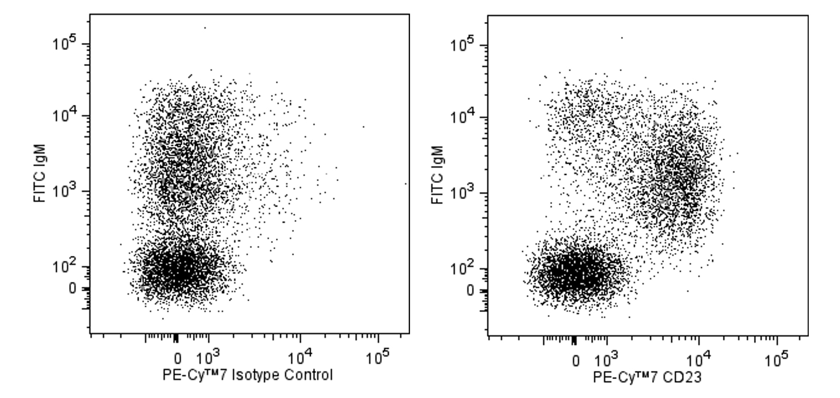
The B3B4 monoclonal antibody specifically binds to CD23, the low affinity IgE Fc receptor (FcεRII) expressed on mature resting conventional B lymphocytes, but not on B-1 cells (CD5+ B cells) or T lymphocytes. It does not react with high-affinity IgE receptors, as demonstrated on mouse mast cell lines. The regulation of CD23 surface expression on activated B cells appears to be complex, depending upon the mode of activation and the presence of cytokines. IgE synthesis is negatively regulated by CD23, and CD23 expression is upregulated on splenocytes in the presence of IgE. CD23 is also upregulated on follicular dendritic cells in the lymph nodes of immunized mice, and a subset of splenic dendritic cells expresses CD23. The B3B4 antibody abrogates antigen-specific IgE-dependent modulation of immune responses in normal mice. This monoclonal antibody also blocks IgE binding and eosinophil infiltration in the lung of immunized mice. Different in vivo results have been obtained when using the intact B3B4 antibody or the F(ab')2 fragments. B3B4 mAb does not cross-react with rat or human IgE Fc Receptor.

Development References (14)
-
Conrad DH, Waldschmidt TJ, Lee WT, et al. Effect of B cell stimulatory factor-1 (interleukin 4) on Fc epsilon and Fc gamma receptor expression on murine B lymphocytes and B cell lines. J Immunol. 1987; 139(7):2290-2296. (Clone-specific: Flow cytometry, Functional assay, Immunoaffinity chromatography, Immunoprecipitation, Radioimmunoassay). View Reference
-
Coyle AJ, Wagner K, Bertrand C, Tsuyuki S, Bews J, Heusser C. Central role of immunoglobulin (Ig) E in the induction of lung eosinophil infiltration and T helper 2 cell cytokine production: inhibition by a non-anaphylactogenic anti-IgE antibody. J Exp Med. 1996; 183(4):1303-1310. (Clone-specific: Blocking). View Reference
-
Dasic G, Juillard P, Graber P, et al. Critical role of CD23 in allergen-induced bronchoconstriction in a murine model of allergic asthma. Eur J Immunol. 1999; 29(9):2957-2967. (Clone-specific: Blocking, In vivo exacerbation). View Reference
-
Kisselgof AB, Oettgen HC. The expression of murine B cell CD23, in vivo, is regulated by its ligand, IgE. Int Immunol. 1998; 10(9):1377-1384. (Clone-specific: Flow cytometry). View Reference
-
Maeda K, Burton GF, Padgett DA, et al. Murine follicular dendritic cells and low affinity Fc receptors for IgE (Fc epsilon RII). J Immunol. 1992; 148(8):2340-2347. (Biology: Electron microscopy, Immunohistochemistry). View Reference
-
Oshiba A, Hamelmann E, Haczku A, et al. Modulation of antigen-induced B and T cell responses by antigen-specific IgE antibodies. J Immunol. 1997; 159(8):4056-4063. (Clone-specific: Blocking). View Reference
-
Pulendran B, Lingappa J, Kennedy MK, et al. Developmental pathways of dendritic cells in vivo: distinct function, phenotype, and localization of dendritic cell subsets in FLT3 ligand-treated mice. J Immunol. 1997; 159(5):2222-2231. (Clone-specific: Flow cytometry). View Reference
-
Rabin E, Cong YZ, Wortis HH. Loss of CD23 is a consequence of B-cell activation. Implications for the analysis of B-cell lineages. Ann N Y Acad Sci. 1992; 651:130-142. (Clone-specific: Flow cytometry). View Reference
-
Rao M, Lee WT, Conrad DH. Characterization of a monoclonal antibody directed against the murine B lymphocyte receptor for IgE. J Immunol. 1987; 138(6):1845-1851. (Immunogen: Blocking, Immunoprecipitation, Inhibition, Radioimmunoassay). View Reference
-
Stief A, Texido G, Sansig G, et al. Mice deficient in CD23 reveal its modulatory role in IgE production but no role in T and B cell development. J Immunol. 1994; 152(7):3378-3390. (Clone-specific: Flow cytometry, Immunohistochemistry). View Reference
-
Waldschmidt T, Snapp K, Foy T, Tygrett L, Carpenter C. B-cell subsets defined by the Fc epsilon R. Ann N Y Acad Sci. 1992; 651:84-98. (Biology). View Reference
-
Waldschmidt TJ, Conrad DH, Lynch RG. Expression of B cell surface receptors. II. IL-4 can accelerate the developmental expression of the murine B cell IgE Fc receptor. J Immunol. 1989; 143(9):2820-2827. (Clone-specific: Flow cytometry). View Reference
-
Waldschmidt TJ, Conrad DH, Lynch RG. The expression of B cell surface receptors. I. The ontogeny and distribution of the murine B cell IgE Fc receptor. J Immunol. 1988; 140(7):2148-2154. (Clone-specific: Flow cytometry, Immunoaffinity chromatography). View Reference
-
Yu P, Kosco-Vilbois M, Richards M, Kohler G, Lamers MC. Negative feedback regulation of IgE synthesis by murine CD23. Nature. 1994; 369(6483):753-756. (Biology). View Reference
Please refer to Support Documents for Quality Certificates
Global - Refer to manufacturer's instructions for use and related User Manuals and Technical data sheets before using this products as described
Comparisons, where applicable, are made against older BD Technology, manual methods or are general performance claims. Comparisons are not made against non-BD technologies, unless otherwise noted.
For Research Use Only. Not for use in diagnostic or therapeutic procedures.