





.png)


Preparation And Storage


Recommended Assay Procedures
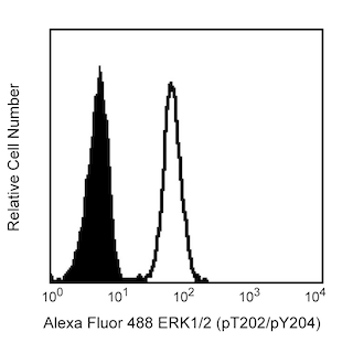
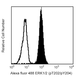
This antibody conjugate is suitable for intracellular staining of human whole blood (using BD Phosflow™ Lyse/Fix Buffer) and peripheral blood mononuclear cells (using BD Cytofix™ Fixation Buffer or BD Phosflow™ Fix Buffer I). Any of the three BD Phosflow™ permeabilization buffers may be used.
This mAb was characterized by flow cytometry (Flow) and western blot analysis (WB) using these model systems:
Method Species Cells Treatment Fixation Perm buffer Result
Flow Human PBMC PMA Fixation Buffer I, II or III Positive Staining
Flow Human Whole Blood PMA Lyse/Fix I, II or III Positive Staining
WB Human A431 Cell Lysate EGF Not Applicable Not Applicable 44/42 kDa
Product Notices
- This reagent has been pre-diluted for use at the recommended Volume per Test. We typically use 1 × 10^6 cells in a 100-µl experimental sample (a test).
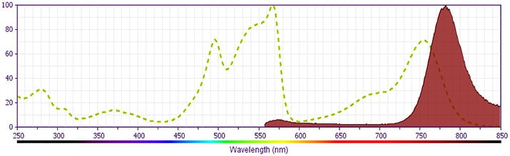
- PE-Cy7 is a tandem fluorochrome composed of R-phycoerythrin (PE), which is excited by 488-nm light and serves as an energy donor, coupled to the cyanine dye Cy7, which acts as an energy acceptor and fluoresces maximally at 780 nm. PE-Cy7 tandem fluorochrome emission is collected in a detector for fluorescence wavelengths of 750 nm and higher. Although every effort is made to minimize the lot-to-lot variation in the efficiency of the fluorochrome energy transfer, differences in the residual emission from PE may be observed. Therefore, we recommend that individual compensation controls be performed for every PE-Cy7 conjugate. PE-Cy7 is optimized for use with a single argon ion laser emitting 488-nm light, and there is no significant overlap between PE-Cy7 and FITC emission spectra. When using dual-laser cytometers, which may directly excite both PE and Cy7, we recommend the use of cross-beam compensation during data acquisition or software compensation during data analysis.
- Cy is a trademark of Amersham Biosciences Limited. This conjugated product is sold under license to the following patents: US Patent Nos. 5,486,616; 5,569,587; 5,569,766; 5,627,027.
- This product is subject to proprietary rights of Amersham Biosciences Corp. and Carnegie Mellon University and made and sold under license from Amersham Biosciences Corp. This product is licensed for sale only for research. It is not licensed for any other use. If you require a commercial license to use this product and do not have one return this material, unopened to BD Biosciences, 10975 Torreyana Rd, San Diego, CA 92121 and any money paid for the material will be refunded.
- Please observe the following precautions: Absorption of visible light can significantly alter the energy transfer occurring in any tandem fluorochrome conjugate; therefore, we recommend that special precautions be taken (such as wrapping vials, tubes, or racks in aluminum foil) to prevent exposure of conjugated reagents, including cells stained with those reagents, to room illumination.
- Caution: Sodium azide yields highly toxic hydrazoic acid under acidic conditions. Dilute azide compounds in running water before discarding to avoid accumulation of potentially explosive deposits in plumbing.
- Source of all serum proteins is from USDA inspected abattoirs located in the United States.
- For fluorochrome spectra and suitable instrument settings, please refer to our Multicolor Flow Cytometry web page at www.bdbiosciences.com/colors.
- Please refer to www.bdbiosciences.com/us/s/resources for technical protocols.
- Warning: Some APC-Cy7 and PE-Cy7 conjugates show changes in their emission spectrum with prolonged exposure to formaldehyde. If you are unable to analyze fixed samples within four hours, we recommend that you use BD™ Stabilizing Fixative (Cat. No. 338036).
Companion Products


The members of the Mitogen-Activated Protein Kinase (MAPK) family are components of a key signal transduction cascade that links events at the cell surface to responses in the nucleus. The signaling cascade is found in species as varied as yeast and humans, with many of the proteins being well conserved. In mammals the most widely studied members of the cascade are the Extracellular signal-Regulated Kinases, ERK1 (p44 MAPK) and ERK2 (p42 MAPK). ERK1 and ERK2 share 85% homology and are activated by extracellular signals such as growth factors, hormones, and phorbol esters. Activation occurs through a series of phosphorylations by kinases activating other kinases and eventually leading to phosphorylation of the ERKs. Growth factor stimulation leads to activation of Ras and Raf, leading to phosphorylation of MEK1 (MAPK/ERK kinase) which, in turn, activates the ERKs via dual phosphorylation. Once activated, the ERKs phosphorylate other cytoplasmic signalling molecules, cell-surface receptors, microtubule-associated proteins, and transcription factors in the nucleus. Thus, the active ERK has myriad downstream effectors that implicate it in the control of cell proliferation and differentiation, as well as regulation of the cytoskeleton. Furthermore, studies have shown that elevated ERK activity is associated with some cancers.
The 20A monoclonal antibody recognizes the phosphorylated threonine 202 and tyrosine 204 (pT202/pY204) of human ERK1 and pT184/pY186 of human ERK2. The orthologous phosphorylation sites in murine ERK1 and ERK2 are T203/Y205 and T183/Y185.
Development References (3)
-
Cobb MH, Boulton TG, Robbins DJ. Extracellular signal-regulated kinases: ERKs in progress. Cell Regul. 1991; 2:965-978. (Biology). View Reference
-
Sivaraman VS, Wang H, Nuovo GJ, Malbon CC. Hyperexpression of mitogen-activated protein kinase in human breast cancer. J Clin Invest. 1997; 99(7):1478-1483. (Biology). View Reference
-
Treisman R. Regulation of transcription by MAP kinase cascades. Curr Opin Cell Biol. 1996; 8:205-215. (Biology). View Reference
Please refer to Support Documents for Quality Certificates
Global - Refer to manufacturer's instructions for use and related User Manuals and Technical data sheets before using this products as described
Comparisons, where applicable, are made against older BD Technology, manual methods or are general performance claims. Comparisons are not made against non-BD technologies, unless otherwise noted.
For Research Use Only. Not for use in diagnostic or therapeutic procedures.
