





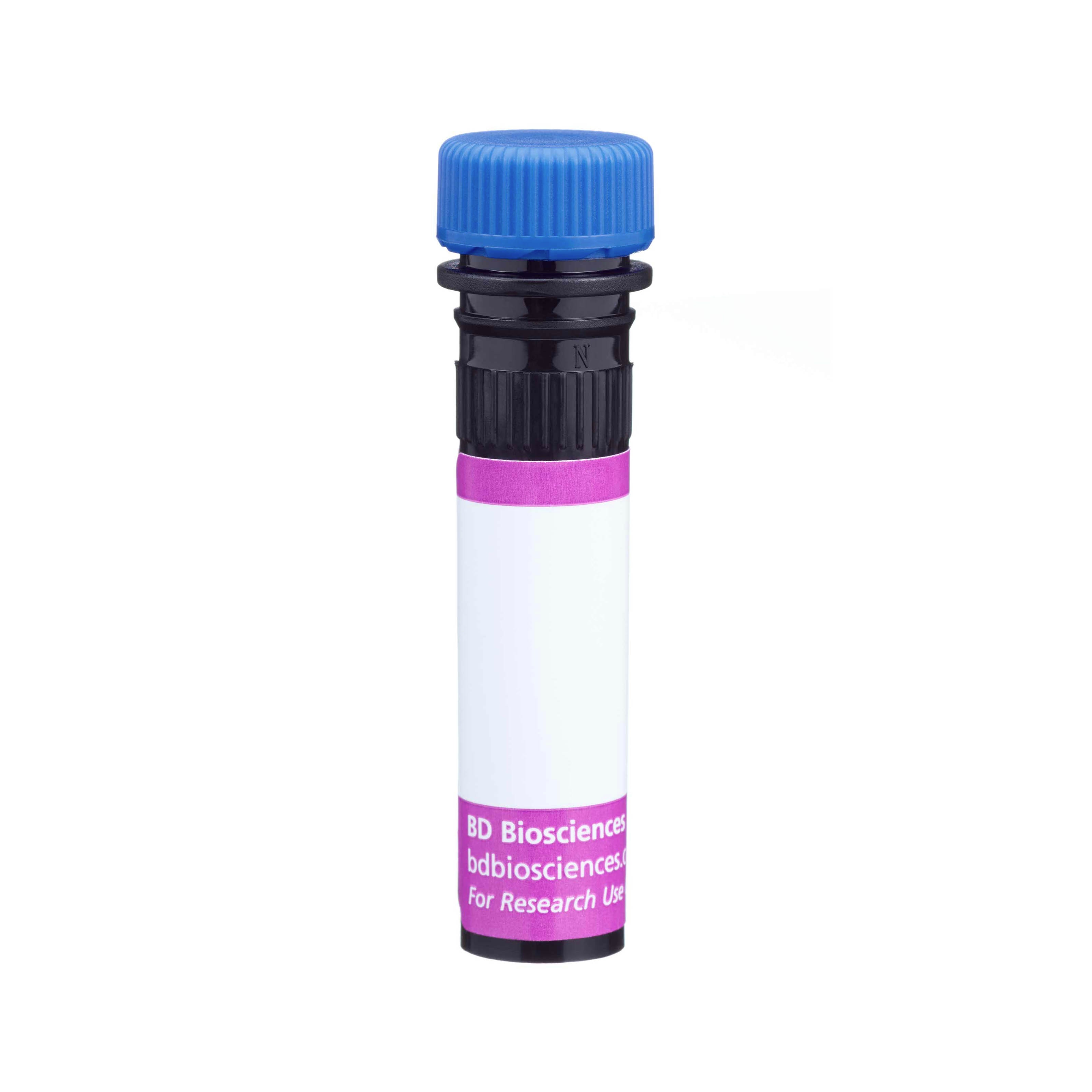


Preparation And Storage


Recommended Assay Procedures
For optimal and reproducible results, BD Horizon Brilliant Stain Buffer should be used anytime two or more BD Horizon Brilliant dyes are used in the same experiment. Fluorescent dye interactions may cause staining artifacts which may affect data interpretation. The BD Horizon Brilliant Stain Buffer was designed to minimize these interactions. More information can be found in the Technical Data Sheet of the BD Horizon Brilliant Stain Buffer (Cat. No. 563794).
Product Notices
- This reagent has been pre-diluted for use at the recommended Volume per Test. We typically use 1 × 10^6 cells in a 100-µl experimental sample (a test).
- Please refer to www.bdbiosciences.com/us/s/resources for technical protocols.
- Pacific Blue™ is a trademark of Molecular Probes, Inc., Eugene, OR.
- Caution: Sodium azide yields highly toxic hydrazoic acid under acidic conditions. Dilute azide compounds in running water before discarding to avoid accumulation of potentially explosive deposits in plumbing.
- For fluorochrome spectra and suitable instrument settings, please refer to our Multicolor Flow Cytometry web page at www.bdbiosciences.com/colors.
- Source of all serum proteins is from USDA inspected abattoirs located in the United States.
- An isotype control should be used at the same concentration as the antibody of interest.
Companion Products




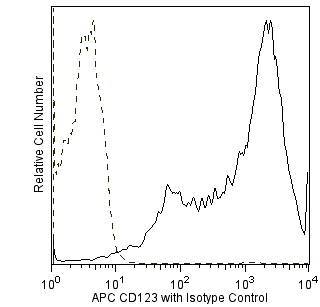
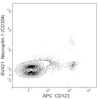
The U21-1283 monoclonal antibody specifically recognizes Neuropilin-1 (NRP1), also known as CD304, Blood dendritic cell antigen 4 (BDCA4), and Vascular endothelial cell growth factor 165 receptor (VEGF165R). CD304 is a type I transmembrane glycoprotein involved in the development of the nervous and cardiovascular systems. It mediates the interaction, growth, survival, and migration of a variety of normal and tumor cells. CD304 is expressed on neurons, thymocytes, regulatory T cells, a subset of T follicular helper cells, dendritic cells, endothelial cells, and certain tumor cells. Neuropilin-1 has a very short cytoplasmic domain and interacts with various coreceptors to form ligand-binding, signal-transducing receptor complexes. CD304 complexes with Plexin-A family members to bind chemorepellent Class 3 Semaphorins and guide neuronal axon growth. It also functions as a coreceptor with VEGFR2/CD309 to stimulate angiogenesis in response to VEGF165. CD304 mediates the interactions between some T cells and dendritic cells.
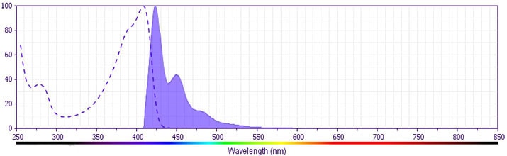
The antibody was conjugated to BD Horizon BV421 which is part of the BD Horizon Brilliant™ Violet family of dyes. With an Ex Max of 407-nm and Em Max at 421-nm, BD Horizon BV421 can be excited by the violet laser and detected in the standard Pacific Blue™ filter set (eg, 450/50-nm filter). BD Horizon BV421 conjugates are very bright, often exhibiting a 10 fold improvement in brightness compared to Pacific Blue conjugates.
Development References (6)
-
Bruder D et al. Neuropilin-1: a surface marker of regulatory T cells.. Eur J Immunol. 2004; 34(3):623-630. (Biology). View Reference
-
Dzionek A et al. BDCA-2, BDCA-3, and BDCA-4: three markers for distinct subsets of dendritic cells in human peripheral blood.. J Immunol. 2000; 165(11):6037-6046. (Biology). View Reference
-
Mizui M, Kumanogoh A, Kikutani H. Immune semaphorins: novel features of neural guidance molecules.. J Clin Immunol. 2009; 29(1):1-11. (Biology). View Reference
-
Pan Q et al. Neuropilin-1 binds to VEGF121 and regulates endothelial cell migration and sprouting.. J Biol Chem. 2007; 282(33):24049-24056. (Biology). View Reference
-
Renand A, Milpied P, Rossignol J, et al. Neuropilin-1 expression characterizes T follicular helper (Tfh) cells activated during B cell differentiation in human secondary lymphoid organs.. PLoS ONE. 2013; 8(12):e85589. (Biology). View Reference
-
Soker S, Takashima S, Miao HQ, Neufeld G, Klagsbrun M. Neuropilin-1 is expressed by endothelial and tumor cells as an isoform-specific receptor for vascular endothelial growth factor.. Cell. 1998; 92(6):735-45. (Biology). View Reference
Please refer to Support Documents for Quality Certificates
Global - Refer to manufacturer's instructions for use and related User Manuals and Technical data sheets before using this products as described
Comparisons, where applicable, are made against older BD Technology, manual methods or are general performance claims. Comparisons are not made against non-BD technologies, unless otherwise noted.
For Research Use Only. Not for use in diagnostic or therapeutic procedures.
