







Preparation And Storage


Recommended Assay Procedures
BD™ CompBeads can be used as surrogates to assess fluorescence spillover (Compensation). When fluorochrome conjugated antibodies are bound to CompBeads, they have spectral properties very similar to cells. However, for some fluorochromes there can be small differences in spectral emissions compared to cells, resulting in spillover values that differ when compared to biological controls. It is strongly recommended that when using a reagent for the first time, users compare the spillover on cells and CompBead to ensure that BD Comp beads are appropriate for your specific cellular application.
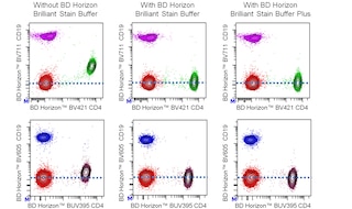
For optimal and reproducible results, BD Horizon Brilliant Stain Buffer should be used anytime two or more BD Horizon Brilliant dyes are used in the same experiment. Fluorescent dye interactions may cause staining artifacts which may affect data interpretation. The BD Horizon Brilliant Stain Buffer was designed to minimize these interactions. More information can be found in the Technical Data Sheet of the BD Horizon Brilliant Stain Buffer (Cat. No. 563794/566349) or the BD Horizon Brilliant Stain Buffer Plus (Cat. No. 566385).
Product Notices
- This reagent has been pre-diluted for use at the recommended Volume per Test. We typically use 1 × 10^6 cells in a 100-µl experimental sample (a test).
- An isotype control should be used at the same concentration as the antibody of interest.
- Caution: Sodium azide yields highly toxic hydrazoic acid under acidic conditions. Dilute azide compounds in running water before discarding to avoid accumulation of potentially explosive deposits in plumbing.
- Source of all serum proteins is from USDA inspected abattoirs located in the United States.
- Pacific Blue™ is a trademark of Molecular Probes, Inc., Eugene, OR.
- For fluorochrome spectra and suitable instrument settings, please refer to our Multicolor Flow Cytometry web page at www.bdbiosciences.com/colors.
- BD Horizon Brilliant Violet 421 is covered by one or more of the following US patents: 8,158,444; 8,362,193; 8,575,303; 8,354,239.
- BD Horizon Brilliant Stain Buffer is covered by one or more of the following US patents: 8,110,673; 8,158,444; 8,575,303; 8,354,239.
- Please refer to http://regdocs.bd.com to access safety data sheets (SDS).
- Please refer to www.bdbiosciences.com/us/s/resources for technical protocols.
Companion Products




The DCN46 antibody specifically binds to dendritic cell-specific ICAM-3 grabbing nonintegrin (DC-SIGN or CD209), a type-II membrane protein of approximately 44 kDa with a mannose-binding C-type lectin domain. It is highly expressed on dendritic cells in mucosal tissues. Its sequence is identical to the HIV-1 envelope gp120-binding C-type lectin, and reports suggest that DC-SIGN binds to HIV-1 gp120 and effectively transmits infectious HIV-1 to resting T lymphocytes expressing CD4 and chemokine receptors. The C-type lectin domain of DC-SIGN is also capable of binding other pathogenic viruses, bacteria, and parasites. Reports also suggest that DC-SIGN enables the highly efficient migration of dendritic cells from blood into the tissues. It can interact with ICAM-2, which has a similar sequence as ICAM-3, and is abundantly expressed on vascular and lymphoid endothelium. Thus, DC-SIGN mediates dendritic cells rolling and transendothelial migration, and its interaction with ICAM-2 is essential to specific migratory functions of dendritic cells.
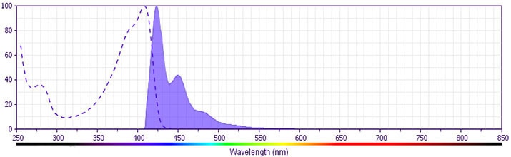
The antibody was conjugated to BD Horizon BV421 which is part of the BD Horizon Brilliant™ Violet family of dyes. With an Ex Max near 407 nm and Em Max near 421 nm, BD Horizon BV421 can be excited by the violet laser (405 nm) and detected with a 450/50 nm filter. BD Horizon BV421 conjugates are very bright, often exhibiting a 10 fold improvement in brightness compared to Pacific BlueTM conjugates. Due to nearly identical excitation and emission properties but different spillover characteristics, BD Horizon BV421, Pacific Blue, and BD Horizon V450 cannot be used simultaneously.
Development References (5)
-
Appelmelk BJ, van Die I, van Vliet SJ, et al. Carbohydrate profiling identifies new pathogens that interact with dendritic cell-specific ICAM-3-grabbing nonintegrin on dendritic cells. J Immunol. 2003; 170(4):1635-1639. (Biology). View Reference
-
Gruber A, Chalmers AS, Popov S, Ruprecht RM. Functional aspects of binding of monoclonal antibody DCN46 to DC-SIGN on dendritic cells.. Immunol Lett. 2002; 84(2):103-8. (Clone-specific: Flow cytometry, Fluorescence activated cell sorting, Functional assay). View Reference
-
Sallusto F, Cella M, Danieli C, Lanzavecchia A. Dendritic cells use macropinocytosis and the mannose receptor to concentrate macromolecules in the major histocompatibility complex class II compartment: downregulation by cytokines and bacterial products. J Exp Med. 1995; 182(2):389-400. (Biology). View Reference
-
Steinman RM, Granelli-Piperno A, Pope M, et al. The interaction of immunodeficiency viruses with dendritic cells. Curr Top Microbiol Immunol. 2003; 276:1-30. (Biology). View Reference
-
Steinman RM. DC-SIGN: a guide to some mysteries to dendritic cells. Cell. 2000; 100(5):491-494. (Biology). View Reference
Please refer to Support Documents for Quality Certificates
Global - Refer to manufacturer's instructions for use and related User Manuals and Technical data sheets before using this products as described
Comparisons, where applicable, are made against older BD Technology, manual methods or are general performance claims. Comparisons are not made against non-BD technologies, unless otherwise noted.
For Research Use Only. Not for use in diagnostic or therapeutic procedures.
