-
Your selected country is
Switzerland
- Change country/language
Old Browser
This page has been recently translated and is available in French now.
Looks like you're visiting us from {countryName}.
Would you like to stay on the current country site or be switched to your country?
Multiparameter flow cytometric analysis of CD3 expression on Human peripheral blood leukocyte populations. Human whole blood was stained with either BD Horizon™ BUV563 Mouse IgG1, κ Isotype Control (Cat. No. 612920; Left Plot) or BD Horizon™ BUV563 Mouse Anti-Human CD3 antibody (Cat. No. 569789; Right Plot) at 0.5 μg/test. Erythrocytes were lysed with BD FACS™ Lysing Solution (Cat. No. 349202). The bivariate pseudocolor density plot showing the correlated expression of CD3 (or Ig Isotype control staining) versus side light-scatter (SSC-A) signals was derived from gated events with the forward and side light-scatter characteristics of intact leukocytes. Flow cytometry and data analysis were performed using a BD LSRFortessa™X-20 Cell Analyzer System and FlowJo™ Software. Data shown on this Technical Data Sheet are not lot specific.
BD Horizon™ BUV563 Mouse Anti-Human CD3
Regulatory Status Legend
Any use of products other than the permitted use without the express written authorization of Becton, Dickinson and Company is strictly prohibited.
Preparation And Storage
Recommended Assay Procedures
BD® CompBeads can be used as surrogates to assess fluorescence spillover (compensation). When fluorochrome-conjugated antibodies are bound to BD® CompBeads, they have spectral properties very similar to cells. However, for some fluorochromes there can be small differences in spectral emissions compared to cells, resulting in spillover values that differ when compared to biological controls. It is strongly recommended that when using a reagent for the first time, users compare the spillover on cells and BD® CompBeads. This will ensure that BD® CompBeads are appropriate for your specific cellular application.
For optimal and reproducible results, BD Horizon Brilliant™ Stain Buffer should be used anytime BD Horizon Brilliant dyes are used in a multicolor flow cytometry panel. Fluorescent dye interactions may cause staining artifacts which may affect data interpretation. The BD Horizon Brilliant Stain Buffer was designed to minimize these interactions. When BD Horizon Brilliant Stain Buffer is used in the multicolor panel, it should also be used in the corresponding compensation controls for all dyes to achieve the most accurate compensation. For the most accurate compensation, compensation controls created with either cells or beads should be exposed to BD Horizon Brilliant Stain Buffer for the same length of time as the corresponding multicolor panel. More information can be found in the Technical Data Sheet of the BD Horizon Brilliant Stain Buffer (Cat. No. 563794/566349) or the BD Horizon Brilliant Stain Buffer Plus (Cat. No. 566385).
Note: When using high concentrations of antibody, background binding of this dye to erythroid cell subsets (mature erythrocytes and precursors) has been observed. For researchers studying these cell populations, or in cases where light scatter gating does not adequately exclude these cells from the analysis, this background may be an important factor to consider when selecting reagents for panel(s).
Product Notices
- Please refer to www.bdbiosciences.com/us/s/resources for technical protocols.
- Since applications vary, each investigator should titrate the reagent to obtain optimal results.
- An isotype control should be used at the same concentration as the antibody of interest.
- Caution: Sodium azide yields highly toxic hydrazoic acid under acidic conditions. Dilute azide compounds in running water before discarding to avoid accumulation of potentially explosive deposits in plumbing.
- For fluorochrome spectra and suitable instrument settings, please refer to our Multicolor Flow Cytometry web page at www.bdbiosciences.com/colors.
- Human donor specific background has been observed in relation to the presence of anti-polyethylene glycol (PEG) antibodies, developed as a result of certain vaccines containing PEG, including some COVID-19 vaccines. We recommend use of BD Horizon Brilliant™ Stain Buffer in your experiments to help mitigate potential background. For more information visit https://www.bdbiosciences.com/en-us/support/product-notices.
- Please refer to http://regdocs.bd.com to access safety data sheets (SDS).
- For U.S. patents that may apply, see bd.com/patents.
Companion Products
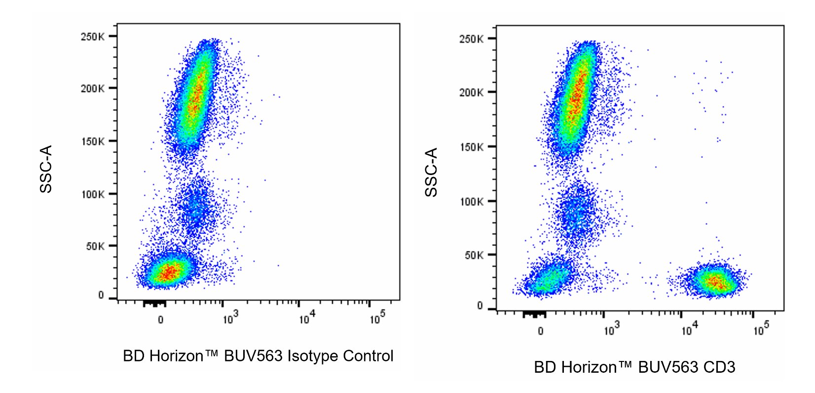
The UCHT1 monoclonal antibody specifically binds to the human CD3ε-chain, a 20-kDa subunit of the CD3/T cell antigen receptor complex. CD3ε is expressed on 70-80% of normal human peripheral blood lymphocytes and 60-85% of thymocytes. Studies from the HLDA Workshop show that this antibody is mitogenic for CD3ε-positive cells when used in conjunction with costimulatory agents such as pokeweed mitogen or anti-CD28 antibody. CD3 plays a central role in signal transduction during antigen recognition. The UCHT1 antibody stains both surface and intracellular CD3ε unlike the other CD3 clone, HIT3a, that stains only extracellular CD3ε.
Development References (9)
-
Balasko A, Graydon C, Fowke KR. Novel in vitro invariant natural killer T cell functional assays.. J Immunol Methods. 2021; 499:113171. (Clone-specific: Flow cytometry). View Reference
-
Baracho GV, Kara N, Rigaud S, Lo E, Widmann SJ, Tyznik AJ. Functional phenotyping of circulating human cytotoxic T cells and NK cells using a 16-color flow cytometry panel.. STAR Protoc. 2022; 3(1):101069. (Clone-specific: Cytotoxicity, Flow cytometry, Functional assay). View Reference
-
Beverley PC, Callard RE. Distinctive functional characteristics of human "T" lymphocytes defined by E rosetting or a monoclonal anti-T cell antibody. Eur J Immunol. 1981; 11(4):329-334. (Immunogen: Flow cytometry, Fluorescence activated cell sorting). View Reference
-
Burns GF, Boyd AW, Beverley PC. Two monoclonal anti-human T lymphocyte antibodies have similar biologic effects and recognize the same cell surface antigen. J Immunol. 1982; 129(4):1451-1457. (Clone-specific: Blocking, Functional assay, Immunoprecipitation, Inhibition, Radioimmunoassay). View Reference
-
Ernst DN, Shih CC. CD3 complex. J Biol Regul Homeost Agents. 2000; 14(3):226-229. (Biology). View Reference
-
Knapp W. W. Knapp .. et al., ed. Leucocyte typing IV : white cell differentiation antigens. Oxford New York: Oxford University Press; 1989:1-1182.
-
McMichael AJ. A.J. McMichael .. et al., ed. Leucocyte typing III : white cell differentiation antigens. Oxford New York: Oxford University Press; 1987:1-1050.
-
Van Wauwe JP, Goossens JG, Beverley PC. Human T lymphocyte activation by monoclonal antibodies; OKT3, but not UCHT1, triggers mitogenesis via an interleukin 2-dependent mechanism. J Immunol. 1984; 133(1):129-132. (Clone-specific: Flow cytometry, Functional assay, Stimulation). View Reference
-
Wang SR, Zhong N, Zhang XM, et al. OMIP 071: A 31-Parameter Flow Cytometry Panel for In-Depth Immunophenotyping of Human T-Cell Subsets Using Surface Markers.. Cytometry A. 2021; 99(3):273-277. (Clone-specific: Flow cytometry). View Reference
Please refer to Support Documents for Quality Certificates
Global - Refer to manufacturer's instructions for use and related User Manuals and Technical data sheets before using this products as described
Comparisons, where applicable, are made against older BD Technology, manual methods or are general performance claims. Comparisons are not made against non-BD technologies, unless otherwise noted.
For Research Use Only. Not for use in diagnostic or therapeutic procedures.
Report a Site Issue
This form is intended to help us improve our website experience. For other support, please visit our Contact Us page.