





Superior Alternative to PerCP-Cy5.5 and BD Horizon Brilliant™ Blue 700 (BB700) Reagents
BD Horizon RealBlue™ 705 (RB705) Reagents leverage an innovative laser-specific fluorochrome, excited primarily by the 488-nm blue laser to offer:
- Minimal cross-laser excitation off the 561-nm yellow-green laser
- A bright fluorochrome to support the detection of low-expression surface and intracellular markers
- Reduced background compared to BB700 with no additional wash recommended
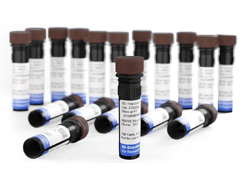

RB705 Reagents Can Be Used Instead of PerCP-Cy5.5 or BB700 on Conventional Flow Cytometers or with PerCP-Cy5.5 or BB700 on Spectral Flow Cytometers
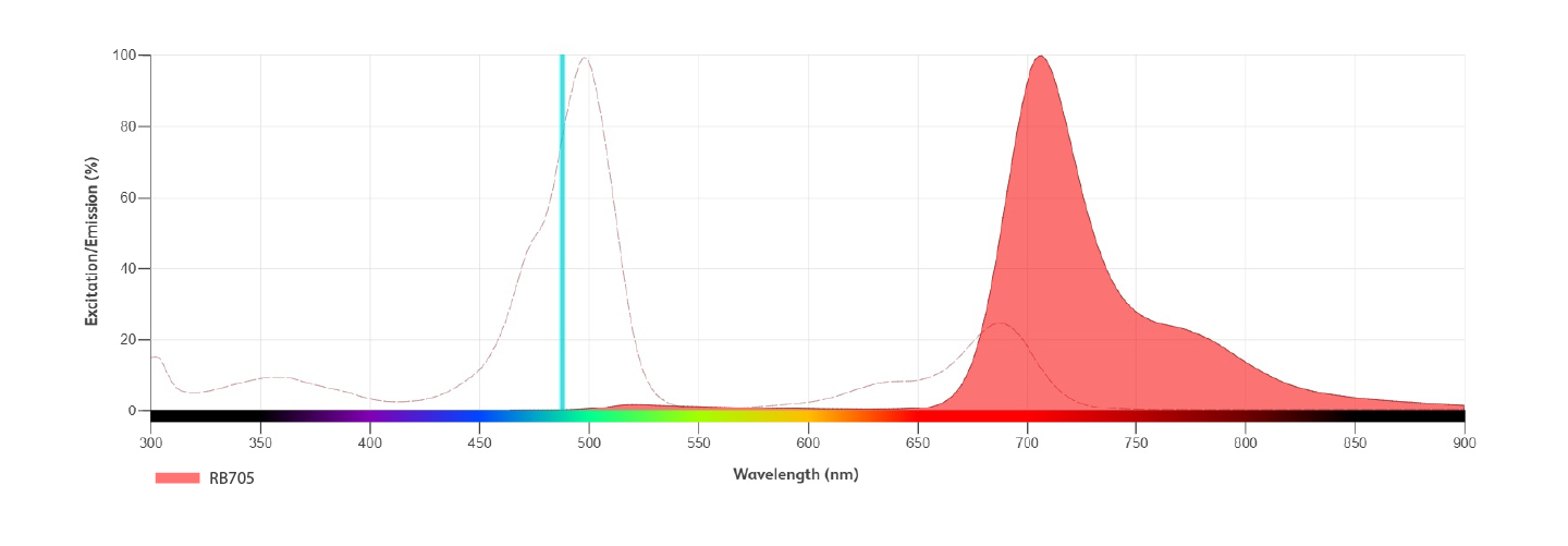
| Format | Ex Max | Em Max | Spectral | Conventional | Relative Brightness | Spillover* (1 = low, 4 = high) | Alternative To |
|---|---|---|---|---|---|---|---|
| RB705 | 498 nm | 707 nm | ✓ | ✓ | 2 | PerCP-Cy5.5, BB700, StarBright™ Blue 700, NovaFluor™ Blue 690, cFluor™ B690 |
*Value may vary based on instrument configuration and settings. Spillover ranking is based on cross-laser excitation on five-laser spectral instruments and does not take into account spillover into adjacent detectors.
Performance
BD Horizon RealBlue™ 705 Reagents Provide Minimal Cross-laser Excitation Off the 561-nm Yellow-green Laser
RB705 has reduced cross-laser excitation
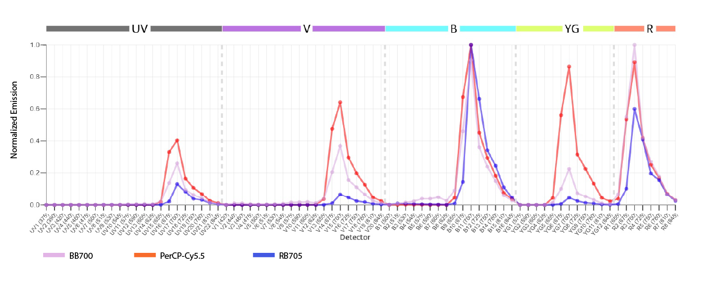
Normalized emission profiles of RB705, BB700 and PerCP-Cy5.5 fluorochromes. Human CD4 SK3 antibody acquired on a BD FACSDiscover™ S8 Cell Sorter.
Compared to PerCP-Cy5.5 and BB700, RB705 is a brighter fluorochrome and has lower spillover spread into the corresponding yellow-green detector
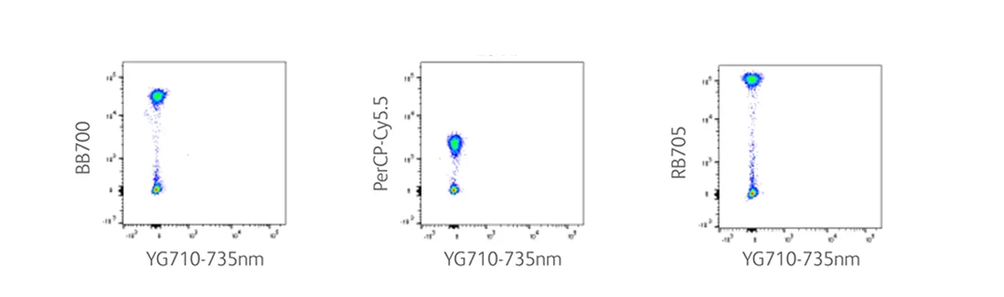
Human whole blood was stained with BD Horizon™ BB700 Reagent, PerCP-Cy5.5 or RB705 Human CD4 (clone SK3), acquired on a BD FACSymphony™ A5 SE Cell Analyzer and compensated with FlowJo™ Software. Note that spillover spread is a function of reagent brightness and antigen density, similar spillover spreading observed at higher brightness indicates lower spillover spread.
Applications
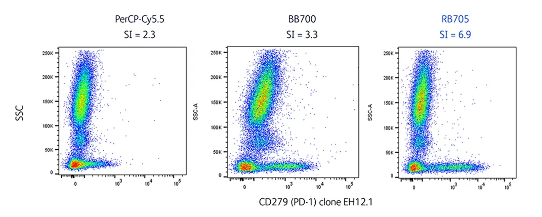
Human whole blood was stained with PerCP-Cy5.5, BB700 or BD Horizon™ RB705 Reagent (right) CD279 (EH12.1) and costained with BD Horizon™ BV421 Reagent CD3 (UCHT1, not shown) followed by acquisition on a BD FACSymphony™ A5 SE Cell Analyzer with compensation in FlowJo™ Software.
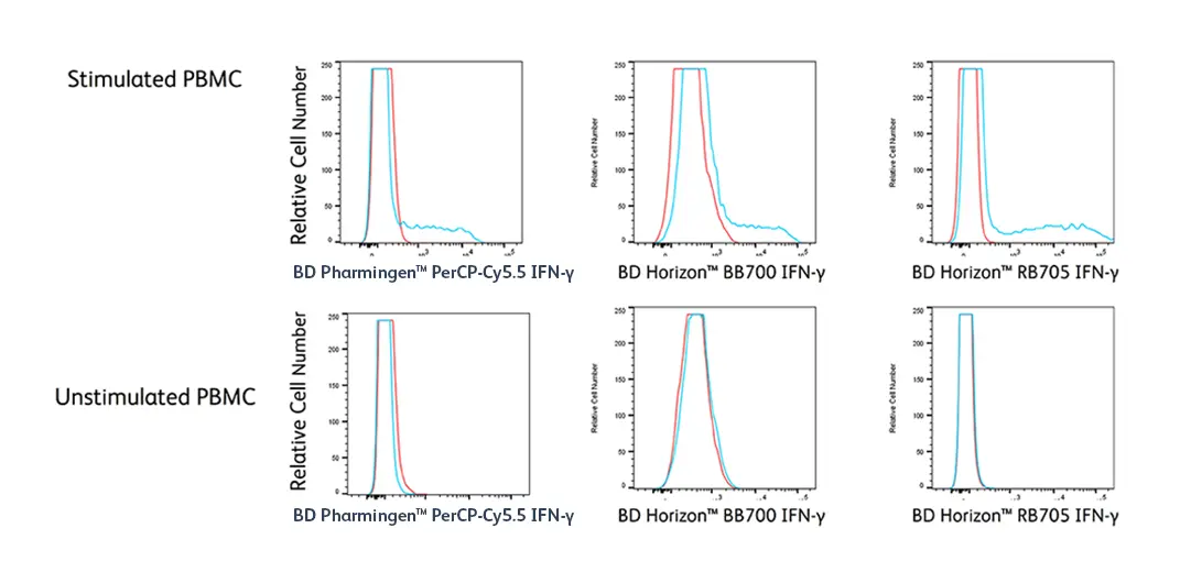
Human PBMCs were stimulated with PMA and ionomycin in the presence of BD GolgiStop™ Protein Transport Inhibitor for 5 hours (top) or left unstimulated (bottom) and then stained with either PerCP-Cy5.5, BB700 or BD Horizon™ RB705 Reagent IFN-γ (clone B27) and matched isotype controls. Samples were acquired on a BD FACSymphony™ A5 SE Cell Analyzer and analyzed with FlowJo™ Software.
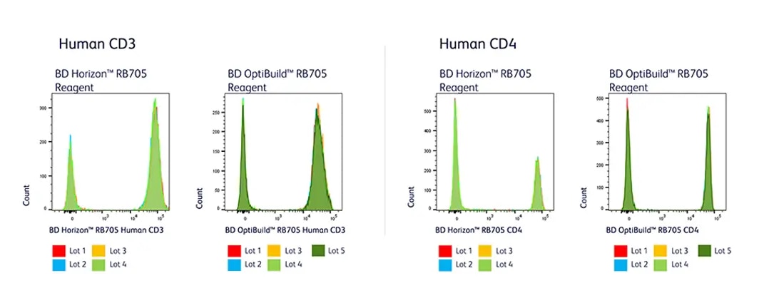
BD Horizon™ RB705 Reagents show stable performance with lot-to-lot consistency across made-to-stock reagents and BD OptiBuild™ On-Demand Reagents. Human whole blood was stained with human CD3 (UCHT1, left) or CD4 (SK3, right) RB705 using several different dye lots, followed by lysis with BD FACS™ Lysing Solution. All specificities were acquired on a BD FACSymphony™ A5 SE Cell Analyzer.
Multicolor Flow Cytometry
BD Horizon™ RB705 Reagents Resolve Well in a 12-color T Cell Inhibitory Panel Acquired on the BD FACSLyric™ Cell Analyzer
Gating scheme of T cell subsets
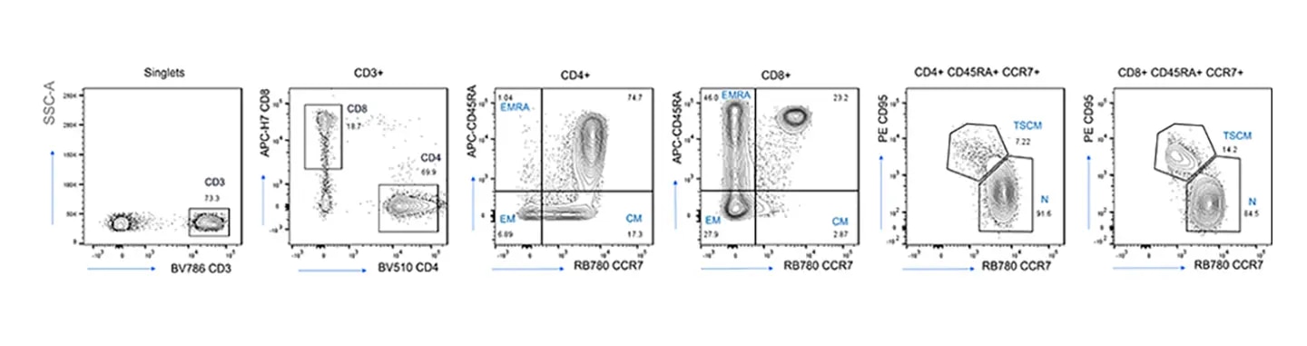
Healthy PBMC were stained with a 12-color inhibitory T cell panel and acquired on the BD FACSLyric™ Flow Cytometry System. Labels on top of each plot show the parental gate from which plots are derived; numbers in each quadrant represent the frequency of cell population; T cell subsets are identified as N, naïve; CM, central memory; EM, effector memory; EMRA, effector memory RA+; and SCM, stem cell like memory.
BD Horizon™ RB705 Reagents resolve low expression surface markers
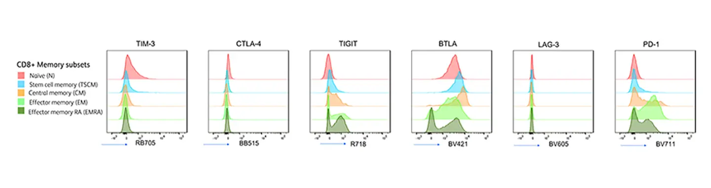
Histogram overlays depict expression pattern of inhibitory receptors TIM-3, CTLA-4, TIGIT, BTLA, LAG-3 and PD-1 on CD8+ T naïve, stem cell memory, central memory, effector memory and effector memory RA cell subsets.
T cell subsets after activation
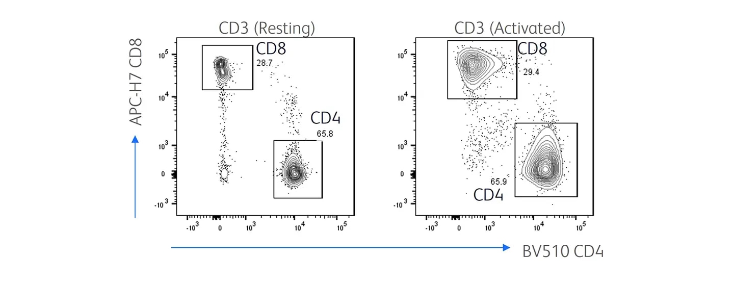
Bivariate plots show CD4/CD8 T cell population derived from activated T cells on Day 3 following immunostaining by a 12-color RB705 panel. Control T cells are from the same donor subjected to similar culture conditions but without activation. Samples were acquired on a 3-laser BD FACSLyric™ Flow Cytometry System.
T cell inhibitory markers are upregulated upon activation
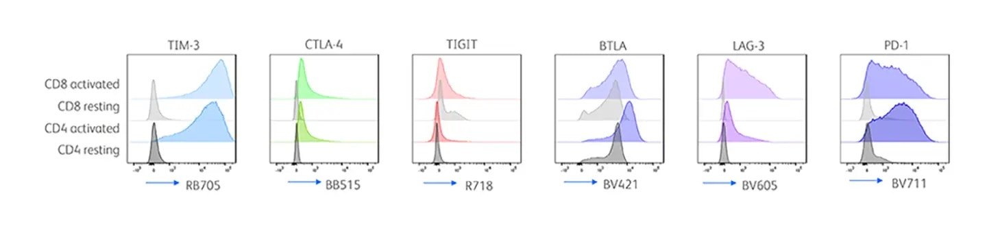
Histogram overlays show expression of T cell inhibitory markers derived from CD4+ resting T cells (deep gray shade) or CD4 activated T cells (deep colored), CD8+ resting T cells (light gray) CD8+ activated T cells (light colored) stained with a 12-color RB705 panel.
BD Horizon™ RB705 Reagents Have Excellent Resolution as Demonstrated in a 17-color Spectral Flow Cytometry Panel Acquired on the BD FACSymphony™ A5 SE Cell Analyzer
Gating strategy for detection of T cell subsets in activated T cells
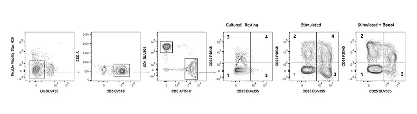
Peripheral blood mononuclear cells were isolated and loaded with Violet Proliferation Dye 450 before stimulation with or without staphylococcal enterotoxin B (SEB, 1 µg/mL) and CD28 (1 µg/mL) for 3 days. PMA (10 ng/mL) and ionomycin (1 µg/mL) were added to the Stimulated + Boost group 4 hours before collecting cells for staining. Cells were then stained with Fixable Viability Stain 620 and antibodies against cell surface markers prior to fixing and permeabilizing with BD Cytofix/Cytoperm™ Fixation/Permeabilization Buffer. Fixed and permeabilized cells were then stained with intracellular antibodies. Stained cells were analyzed on a BD FACSymphony™ A5 SE Cell Analyzer. Gating strategy for detection of T cell subsets after exclusion of doublets, dead cells and lineage-positive cells. CD3+ T cells were selected and divided into CD4+ or CD8+ cells. CD8+ T cells were further evaluated for activation stage based on their expression of CD25 and CD69. Samples were acquired on a BD FACSymphony™ A5 SE Cell Analyzer and analyzed with FlowJo™ Software.
Expression of inhibitory receptors at different activation levels of CD8+ T cells
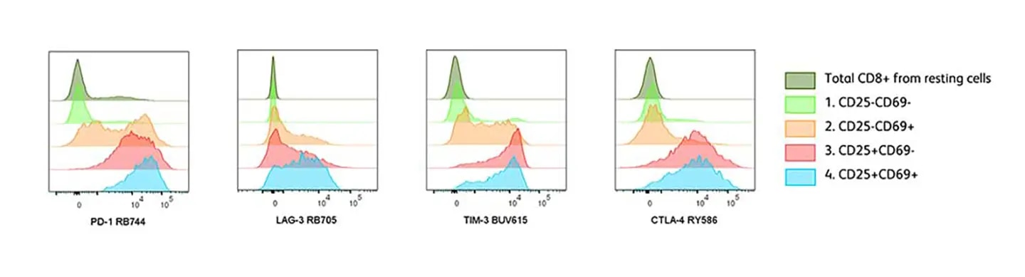
Histogram overlays showing expression of inhibitory receptors on CD8+ T cell subsets from the Stimulated group. Total CD8+ T cells from the unstimulated group (top, dark green).
Cytokines expression at different activation stages of CD8+ T cells
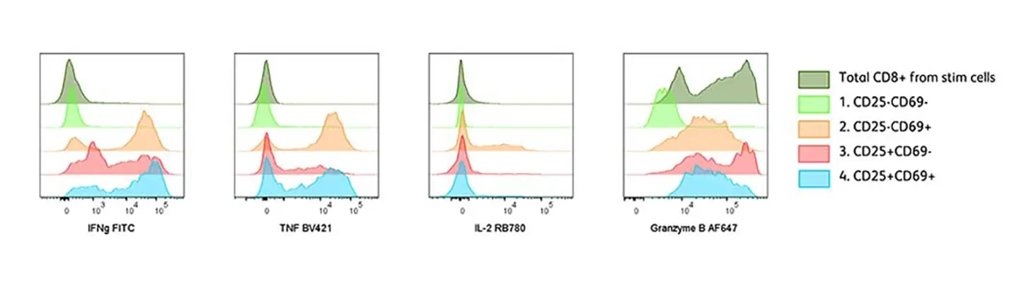
Histogram overlays showing expression of cytokines on CD8+ T cell subsets from the Stimulated + Boost group. Total CD8+ T cells from the unstimulated group (top, dark green).
Fluorochrome marker assignment for a 17-color spectral flow cytometry panel
| Specificity | Clone | Fluorochrome | |
|---|---|---|---|
| UV 355 nm | CD25 | M-A251 | |
CD56 | NCAM16.2 | ||
CD20 | L27 | ||
CD19 | SJ25C1 | ||
CD16 | 3G8 | ||
CD14 | M5E2 | ||
AutoF | N/A | N/A | |
TIM-3 | 7D3 | ||
CD4 | SK3 | ||
| Violet 405 nm | TNF | MAb11 | |
VPD450 | N/A | N/A | |
CD3 | UCHT1 | ||
CD134 | ACT35 | ||
| Blue 488 nm | IFNγ | B27 | |
CD69 | FN50 | ||
FVS620 | N/A | N/A | |
LAG-3 | T47-530 | ||
PD-1 | EH12.1 | ||
II-2 | MQ1-17H12 | ||
| Yellow-Green 561 nm | CTLA-4 | BNI3 | |
| Red 640 nm | GranzB | GB11 | |
CD8 | SK1 |
Fluorochrome marker assignment for a 17-color spectral flow cytometry panel acquired on the BD FACSymphony™ A5 SE Cell Analyzer.
Buffer Compatibility
BD Horizon RealBlue™ 705 Reagents Are Compatible with a Broad Range of Fixation and Permeabilization Systems
| Buffers | Results |
| BD FACS™ Lysing Solution and BD Pharm Lyse™ Lysing Buffer | Compatible |
| CellBox™ Blocking Buffer | Compatible |
| BD Cytofix™ Fixation Buffer | Stable at least 24 hours |
| 1% PFA | Stable at least 24 hours |
| BD Cytofix/Cytoperm™ Fixation and Permeabilization Solution | Compatible with antibody staining before and after fixation |
| BD FACS™ Permeabilizing Solution 2 | Compatible with antibody staining before and after fixation |
| BD Phosflow™ Perm Buffer III | Compatible with antibody staining before and after fixation |
| EDTA and Heparin | Compatible |
| BD Horizon™ Brilliant Stain Buffer (BSB) | Compatible |
-
Brochures
(2)


-
Poster
(1)
-
Performance Guide and Chart
(2)
BD flow cytometers are Class 1 Laser Products. For Research Use Only. Not for use in diagnostic or therapeutic procedures.
The BD FACSLyric™ Flow Cytometer is for Research Use Only with BD FACSuite™ Application for up to 12 colors. Not for use in diagnostic or therapeutic procedures.
CF is a trademark of Biotium, Inc. Cy is a trademark of Global Life Sciences Solutions Germany GmbH or an affiliate doing business as Cytiva. CellBlox is a trademark of Thermo Fisher Scientific.