


Preparation And Storage
Product Notices
- This reagent has been pre-diluted for use at the recommended Volume per Test. We typically use 1 × 10^6 cells in a 100-µl experimental sample (a test).
- An isotype control should be used at the same concentration as the antibody of interest.
- Caution: Sodium azide yields highly toxic hydrazoic acid under acidic conditions. Dilute azide compounds in running water before discarding to avoid accumulation of potentially explosive deposits in plumbing.
- Source of all serum proteins is from USDA inspected abattoirs located in the United States.
- Pacific Blue™ is a trademark of Molecular Probes, Inc., Eugene, OR.
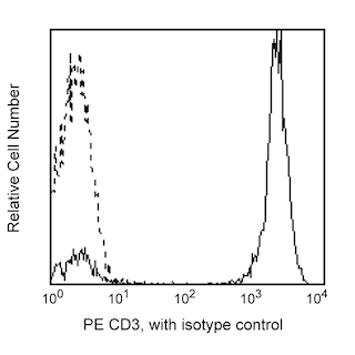
- For fluorochrome spectra and suitable instrument settings, please refer to our Multicolor Flow Cytometry web page at www.bdbiosciences.com/colors.
- Please refer to www.bdbiosciences.com/us/s/resources for technical protocols.
Companion Products


The MIH4 monoclonal antibody specifically binds to CD279, which is also known as, Programmed cell death 1 (PD-1). CD279 is a type I transmembrane glycoprotein that belongs to the Ig superfamily. CD279 is an immunoregulatory receptor that is expressed on expressed on subsets of thymocytes, activated T cells, B cells and myeloid cells. CD279 contains an immunoreceptor tyrosine-based inhibitory motif (ITIM) in its cytoplasmic region. CD273 (PD-L2) and CD274 (PD-L1) are ligands of CD279 and are members of the B7 gene family. Interaction of CD279 with its ligands results in inhibition of T cell proliferation and cytokine secretion. CD279 may play roles in supporting self-tolerance, reducing autoimmunity, or promoting T cell exhaustion associated with certain diseases.
The antibody was conjugated to BD Horizon™ BV421 which is part of the BD Horizon Brilliant™ Violet family of dyes. With an Ex Max of 407-nm and Em Max at 421-nm, BD Horizon BV421 can be excited by the violet laser and detected in the standard Pacific Blue™ filter set (eg, 450/50-nm filter). BD Horizon BV421 conjugates are very bright, often exhibiting a 10 fold improvement in brightness compared to Pacific Blue conjugates.

Development References (6)
-
Bennett F, Luxenberg D, Ling V, et al. Program death-1 engagement upon TCR activation has distinct effects on costimulation and cytokine-driven proliferation: attenuation of ICOS, IL-4, and IL-21, but not CD28, IL-7, and IL-15 responses. J Immunol. 2003; 170(2):711-718. (Biology). View Reference
-
Carter L, Fouser LA, Jussif J, et al. PD-1:PD-L inhibitory pathway affects both CD4(+) and CD8(+) T cells and is overcome by IL-2. Eur J Immunol. 2002; 32:634-643. (Biology). View Reference
-
Freeman GJ, Long AJ, Iwai Y, et al. Engagement of PD-1 immunoinhibitory receptor by a novel B7 family member leads to negative regulation of lymphocyte activation. J Exp Med. 2000; 192:1027-1034. (Biology). View Reference
-
Latchman Y, Wood CR, Chernova T, et al. PD-L2 is a second ligand for PD-1 and inhibits T cell activation. Nat Immunol. 2001; 2(3):261-268. (Biology). View Reference
-
Nishimura H, Minato N, Nakano T, Honjo T. Immunological studies on PD-1 deficient mice: implication of PD-1 as a negative regulator for B cell responses. Int Immunol. 1998; 10(10):1563-1572. (Biology). View Reference
-
Youngnak P, Kozono Y, Kozono H, et al. Differential binding properties of B7-H1 and B7-DC to programmed death-1. Biochem Biophys Res Commun. 2003; 307(3):672-677. (Immunogen: Flow cytometry). View Reference
Please refer to Support Documents for Quality Certificates
Global - Refer to manufacturer's instructions for use and related User Manuals and Technical data sheets before using this products as described
Comparisons, where applicable, are made against older BD Technology, manual methods or are general performance claims. Comparisons are not made against non-BD technologies, unless otherwise noted.
For Research Use Only. Not for use in diagnostic or therapeutic procedures.