Old Browser
This page has been recently translated and is available in French now.
Looks like you're visiting us from {countryName}.
Would you like to stay on the current country site or be switched to your country?


Flow cytometric analysis of Cardiac Troponin T in mouse cardiomyocytes differentiated in vitro. The C2C12 mouse myoblast cell line (ATCC CRL-1772) was cultured for 5 days in low-serum conditions for induction of cell differentiation, fixed with BD Cytofix™ Fixation Buffer (Cat. No. 554655), and permeabilized with BD Perm/Wash™ Buffer (Cat. No. 554723). The cells were stained with either Alexa Fluor® 647 Mouse IgG1, κ Isotype Control (Cat. No. 557732, dashed line) or Alexa Fluor® 647 Mouse Anti-Cardiac Troponin T (solid line). The fluorescence histograms were derived from gated events with the forward and side light-scatter characteristics of intact cells. Flow cytometry was performed on a BD LSR™ II flow cytometer system.

Image analysis of Cardiac Troponin T in mouse heart (view of left atrium). Mouse heart cryosections (5 µm) were fixed with BD Cytofix™ Fixation Buffer (Cat. No. 554655), blocked with 5% goat serum and 1% BSA, permeabilized with 0.2% Triton™ X-100 diluted in PBS, and stained with Alexa Fluor® 647 Mouse Anti-Cardiac Troponin T (pseudocolored red), BD Horizon™ BV480 Rat Anti-Mouse CD31 (Cat. No. 565629, pseudocolored green), and BD Pharmingen™ 7-AAD (Cat. No. 559925, pseudocolored blue). Slides were mounted with ProLong® Gold Antifade Mountant (Life Technologies), and the images and the images were captured on a standard epifluorescence microscope with a 20X objective.


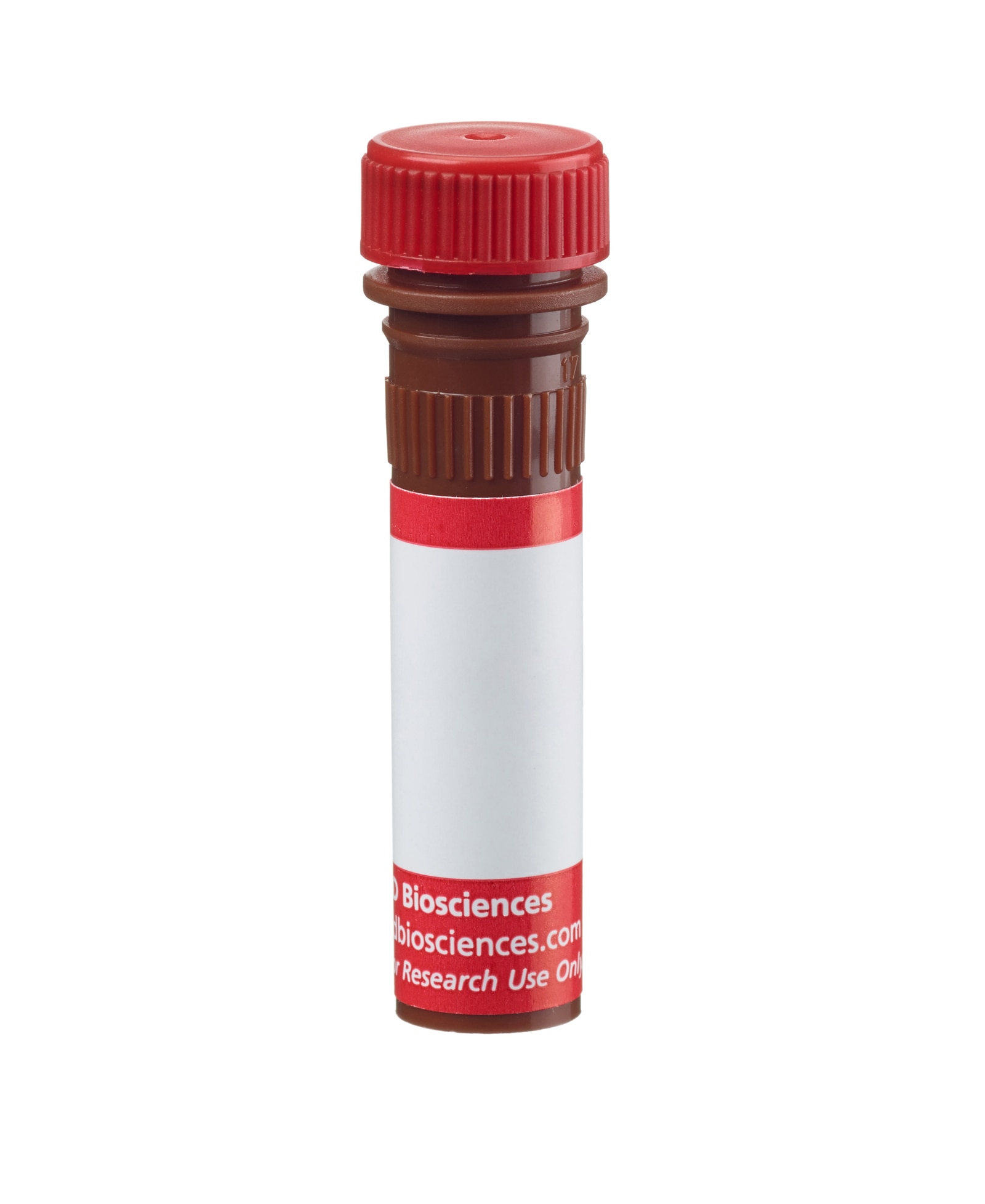
BD Pharmingen™ Alexa Fluor® 647 Mouse Anti-Cardiac Troponin T

BD Pharmingen™ Alexa Fluor® 647 Mouse Anti-Cardiac Troponin T

Regulatory Status Legend
Any use of products other than the permitted use without the express written authorization of Becton, Dickinson and Company is strictly prohibited.
Preparation And Storage
Product Notices
- Since applications vary, each investigator should titrate the reagent to obtain optimal results.
- Please refer to www.bdbiosciences.com/us/s/resources for technical protocols.
- The Alexa Fluor®, Pacific Blue™, and Cascade Blue® dye antibody conjugates in this product are sold under license from Molecular Probes, Inc. for research use only, excluding use in combination with microarrays, or as analyte specific reagents. The Alexa Fluor® dyes (except for Alexa Fluor® 430), Pacific Blue™ dye, and Cascade Blue® dye are covered by pending and issued patents.
- Alexa Fluor® is a registered trademark of Molecular Probes, Inc., Eugene, OR.
- Alexa Fluor® 647 fluorochrome emission is collected at the same instrument settings as for allophycocyanin (APC).
- Caution: Sodium azide yields highly toxic hydrazoic acid under acidic conditions. Dilute azide compounds in running water before discarding to avoid accumulation of potentially explosive deposits in plumbing.
- For fluorochrome spectra and suitable instrument settings, please refer to our Multicolor Flow Cytometry web page at www.bdbiosciences.com/colors.
- An isotype control should be used at the same concentration as the antibody of interest.
- Species cross-reactivity detected in product development may not have been confirmed on every format and/or application.
- Triton is a trademark of the Dow Chemical Company.
- ProLong® is a registered trademark of Thermo Fisher Scientific, Inc. Waltham, MA.
Companion Products


The 13-11 monoclonal antibody specifically recognizes Troponin T type 2 (cardiac), encoded by the gene TNNT2. Troponin T is the tropomyosin-binding subunit of the troponin complex, which also encompasses troponin C and troponin I. This complex regulates muscle contraction in skeletal and cardiac muscle in response to alterations in calcium levels. Troponin T type 2 is solely found in the heart, and genetic alterations in the TNNT2 gene are associated to a series of heart disorders in humans, including hypertrophic cardiomyopathy, dilated cardiomyopathy and left ventricular noncompaction. Cardiac Troponin T can be used as a marker for the identification of cardiomyocytes derived from pluripotent stem cells.
Development References (7)
-
Jáchymová M, Muravská A, Paleček T, et al. Genetic variation screening of TNNT2 gene in a cohort of patients with hypertrophic and dilated cardiomyopathy. Physiol Rev. 2012; 61(2):169-175. (Biology). View Reference
-
Lian X, Hsiao C, Wilson G, et al. Robust cardiomyocyte differentiation from human pluripotent stem cells via temporal modulation of canonical Wnt signaling. Proc Natl Acad Sci U S A. 2012; 109(27):E1848-E1857. (Clone-specific: Flow cytometry). View Reference
-
Malouf NN, McMahon D, Oakeley AE, Anderson PA. A cardiac troponin T epitope conserved across phyla. J Biol Chem. 1992; 267(13):9269-9274. (Immunogen: Electron microscopy, ELISA, Immunofluorescence, Western blot). View Reference
-
Mauritz C, Schwanke K, Reppel M, et al. Generation of functional murine cardiac myocytes from induced pluripotent stem cells. Circulation. 2008; 118(5):507-517. (Clone-specific: Immunofluorescence). View Reference
-
Thierfelder L, Watkins H, MacRae C, et al. Alpha-tropomyosin and cardiac troponin T mutations cause familial hypertrophic cardiomyopathy: a disease of the sarcomere.. Cell. 1994; 77(5):701-12. (Biology). View Reference
-
Uosaki H, Fukushima H, Takeuchi A, et al. Efficient and scalable purification of cardiomyocytes from human embryonic and induced pluripotent stem cells by VCAM1 surface expression. PLoS ONE. 6(8)(Clone-specific: Flow cytometry). View Reference
-
Zhang J, Wilson GF, Soerens AG, et al. Functional cardiomyocytes derived from human induced pluripotent stem cells. Circ Res. 104(4)(Clone-specific: Immunofluorescence). View Reference
Please refer to Support Documents for Quality Certificates
Global - Refer to manufacturer's instructions for use and related User Manuals and Technical data sheets before using this products as described
Comparisons, where applicable, are made against older BD Technology, manual methods or are general performance claims. Comparisons are not made against non-BD technologies, unless otherwise noted.
For Research Use Only. Not for use in diagnostic or therapeutic procedures.
Report a Site Issue
This form is intended to help us improve our website experience. For other support, please visit our Contact Us page.