Old Browser
This page has been recently translated and is available in French now.
Looks like you're visiting us from {countryName}.
Would you like to stay on the current country site or be switched to your country?


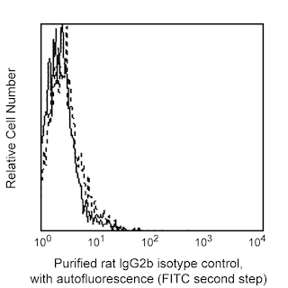
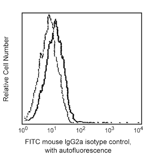
Detection of CXCR5 (CD185) expression on human peripheral lymphocytes by Purified Rat-Anti Human CXCR5 (CD185). Human peripheral blood mononuclear cells were stained with 0.25 µg/test of Purified Rat Anti-Human CXCR5 (CD185) (Cat. No. 552032), using the 3-step staining protocol outlined below, and FITC Mouse Anti-Human CD3 (Cat. No. 555339; left panel) or FITC Mouse Anti-Human CD19 (Cat. No. 555412; right panel). Erythrocytes were lysed with BD Pharm Lyse™ Lysing Buffer (Cat. No. 555899). The data reflects gating on lymphocytes based on forward and side scattered light signals. The level of nonspecific staining was assessed by using Purified Rat IgG2b, ĸ Isotype Control (Cat. No. 555846) and FITC Mouse IgG2a ĸ Isotype Control (Cat. No. 555573), or FITC Mouse IgG1, κ Isotype (Cat. No. 555748). The quadrant markers for the bivariate dot plots were set based on the isotype controls. Flow cytometry was performed using a BD FACScan™ Flow Cytometer (BD Biosciences, San Jose, CA)


BD Pharmingen™ Purifed Rat Anti-Human CXCR5 (CD185)

Regulatory Status Legend
Any use of products other than the permitted use without the express written authorization of Becton, Dickinson and Company is strictly prohibited.
Preparation And Storage
Recommended Assay Procedures
The Purified Rat Anti-Human CXCR5 (CD185) antibody can be used for the immunofluorescent staining and flow cytometric analyses of human leukocytes and cell lines that express CXCR5 (see Figure). A multistep staining procedure is recommended to amplify immunofluorescent signals for the flow cytometric analysis of human CXCR5 expression:
Step 1: Incubate 10e6 cells with 0.06 - 0.5 mg of purified Purified Rat Anti-Human CXCR5 (CD185) antibody at 4°C for 15 - 20 minutes. Wash cells two times with staining medium containing sodium azide (e.g., Dulbecco's PBS or tissue culture medium [without phenol red and biotin] with 0.09% sodium azide and 2% heat-inactivated FCS or 0.2% BSA).
Step 2: Incubate the cells with Biotin Mouse Anti-Rat IgG2b (Cat. No. 553898) at 4°C for 20 minutes. Wash cells two times.
Step 3: Incubate the cells with ≤ 0.06 mg of streptavidin-phycoerythrin (Cat. No. 554061) at 4°C for 20 minutes. Wash two times. Resuspend cells in staining medium and analyze stained cells by flow cyotmetry, using appropriate specificity and compensation controls.
Product Notices
- Since applications vary, each investigator should titrate the reagent to obtain optimal results.
- An isotype control should be used at the same concentration as the antibody of interest.
- Caution: Sodium azide yields highly toxic hydrazoic acid under acidic conditions. Dilute azide compounds in running water before discarding to avoid accumulation of potentially explosive deposits in plumbing.
- Sodium azide is a reversible inhibitor of oxidative metabolism; therefore, antibody preparations containing this preservative agent must not be used in cell cultures nor injected into animals. Sodium azide may be removed by washing stained cells or plate-bound antibody or dialyzing soluble antibody in sodium azide-free buffer. Since endotoxin may also affect the results of functional studies, we recommend the NA/LE (No Azide/Low Endotoxin) antibody format, if available, for in vitro and in vivo use.
- Please refer to www.bdbiosciences.com/us/s/resources for technical protocols.
Companion Products


The RF8B2 monoclonal antibody specifically binds to the human CXC chemokine receptor, CXCR5. CXCR5 (also known as CD185, BLR-1 NLR and MDR15), a seven transmembrane, G-protein-coupled receptor, is the specific receptor for CXC chemokine, CXCL13/BLC/BCA-1. In peripheral blood, CXCR5 expression is restricted to B lymphocytes and a small subset of CD4+ and CD8+ lymphocytes. The restricted expression pattern of CXCR5 on B cells and follicular T helper cells (Tfh) suggests that this receptor functions as a regulator of B and T cell migration. Stimulation of T cells with anti-CD3 monoclonal antibody leads to the down-regulation of CXCR5.
Development References (6)
-
Barella L, Loetscher M, Tobler A, Baggiolini M, Moser B. Sequence variation of a novel heptahelical leucocyte receptor through alternative transcript formation. Biochem J. 1995; 309(3):773-779. (Biology). View Reference
-
Dobner T, Wolf I, Emrich T, Lipp M. Differentiation-specific expression of a novel G protein-coupled receptor from Burkitt's lymphoma. Eur J Immunol. 1992; 22(11):2795-2799. (Biology). View Reference
-
Forster R, Emrich T, Kremmer E, Lipp M. Expression of the G-protein--coupled receptor BLR1 defines mature, recirculating B cells and a subset of T-helper memory cells. Blood. 1994; 84(3):830-840. (Immunogen). View Reference
-
Gunn MD, Ngo VN, Ansel KM, Ekland EH, Cyster JG, Williams LT. A B-cell-homing chemokine made in lymphoid follicles activates Burkitt's lymphoma receptor-1. Nature. 1998; 391(6669):799-803. (Biology). View Reference
-
Kouba M, Vanetti M, Wang X, Schafer M, Hollt V. Cloning of a novel putative G-protein-coupled receptor (NLR) which is expressed in neuronal and lymphatic tissue. FEBS Lett. 1993; 321(2-3):173-178. (Biology). View Reference
-
Legler DF, Loetscher M, Roos RS, Clark-Lewis I, Baggiolini M, Moser B. B cell-attracting chemokine 1, a human CXC chemokine expressed in lymphoid tissues, selectively attracts B lymphocytes via BLR1/CXCR5. J Exp Med. 1998; 187(4):655-660. (Biology). View Reference
Please refer to Support Documents for Quality Certificates
Global - Refer to manufacturer's instructions for use and related User Manuals and Technical data sheets before using this products as described
Comparisons, where applicable, are made against older BD Technology, manual methods or are general performance claims. Comparisons are not made against non-BD technologies, unless otherwise noted.
For Research Use Only. Not for use in diagnostic or therapeutic procedures.
Report a Site Issue
This form is intended to help us improve our website experience. For other support, please visit our Contact Us page.