
Preparation And Storage
Product Notices
- Since applications vary, each investigator should titrate the reagent to obtain optimal results.
- An isotype control should be used at the same concentration as the antibody of interest.
- Caution: Sodium azide yields highly toxic hydrazoic acid under acidic conditions. Dilute azide compounds in running water before discarding to avoid accumulation of potentially explosive deposits in plumbing.
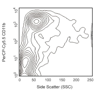
- For fluorochrome spectra and suitable instrument settings, please refer to our Multicolor Flow Cytometry web page at www.bdbiosciences.com/colors.
- Cy is a trademark of GE Healthcare.
- Please refer to www.bdbiosciences.com/us/s/resources for technical protocols.
Companion Products


.png?imwidth=320)
The RMT4-54 monoclonal antibody specifically binds to TIM-4. TIM-4 is encoded by Timd4 (T cell immunoglobulin and mucin domain containing 4). TIM-4 is also known as Spleen, mucin-containing, knockout of lymphotoxin protein (SMUCKLER). TIM-4 is a single-pass type I membrane transmembrane glycoprotein belonging to the TIM family of the immunoglobulin superfamily. TIM-4 is expressed by macrophages and at low levels by dendritic cells. TIM-4 is a phosphatidylserine receptor that enhances the phagocytosis of apoptotic cells. It can serve as a receptor for TIM-1, also known as the Hepatitis A virus cellular receptor 1 (Havcr1).

Development References (3)
-
Curtiss ML, Gorman JV, Businga TR, et al. Tim-1 regulates Th2 responses in an airway hypersensitivity model. Eur J Immunol. 2012; 42(3):651-661. (Clone-specific: Flow cytometry). View Reference
-
Freeman GJ, Casasnovas JM, Umetsu DT, DeKruyff RH. TIM genes: a family of cell surface phosphatidylserine receptors that regulate innate and adaptive immunity.. Immunol Rev. 2010; 235(1):172-89. (Biology). View Reference
-
Nakayama M, Akiba H, Takeda K, et al. Tim-3 mediates phagocytosis of apoptotic cells and cross-presentation. Blood. 2009; 113(16):3821-3830. (Immunogen: Blocking, Flow cytometry, Functional assay, Inhibition, In vivo exacerbation). View Reference
Please refer to Support Documents for Quality Certificates
Global - Refer to manufacturer's instructions for use and related User Manuals and Technical data sheets before using this products as described
Comparisons, where applicable, are made against older BD Technology, manual methods or are general performance claims. Comparisons are not made against non-BD technologies, unless otherwise noted.
For Research Use Only. Not for use in diagnostic or therapeutic procedures.