
Preparation And Storage
Recommended Assay Procedures
BD® CompBeads can be used as surrogates to assess fluorescence spillover (compensation). When fluorochrome conjugated antibodies are bound to BD® CompBeads, they have spectral properties very similar to cells. However, for some fluorochromes there can be small differences in spectral emissions compared to cells, resulting in spillover values that differ when compared to biological controls. It is strongly recommended that when using a reagent for the first time, users compare the spillover on cell and BD® CompBeads to ensure that BD® CompBeads are appropriate for your specific cellular application.
Product Notices
- Please refer to www.bdbiosciences.com/us/s/resources for technical protocols.
- This reagent has been pre-diluted for use at the recommended Volume per Test. We typically use 1 × 10^6 cells in a 100-µl experimental sample (a test).
- An isotype control should be used at the same concentration as the antibody of interest.
- Caution: Sodium azide yields highly toxic hydrazoic acid under acidic conditions. Dilute azide compounds in running water before discarding to avoid accumulation of potentially explosive deposits in plumbing.
- Source of all serum proteins is from USDA inspected abattoirs located in the United States.
- Alexa Fluor® 647 fluorochrome emission is collected at the same instrument settings as for allophycocyanin (APC).
- For fluorochrome spectra and suitable instrument settings, please refer to our Multicolor Flow Cytometry web page at www.bdbiosciences.com/colors.
- This product is provided under an intellectual property license between Life Technologies Corporation and BD Businesses. The purchase of this product conveys to the buyer the non-transferable right to use the purchased amount of the product and components of the product in research conducted by the buyer (whether the buyer is an academic or for-profit entity). The buyer cannot sell or otherwise transfer (a) this product (b) its components or (c) materials made using this product or its components to a third party or otherwise use this product or its components or materials made using this product or its components for Commercial Purposes. Commercial Purposes means any activity by a party for consideration and may include, but is not limited to: (1) use of the product or its components in manufacturing; (2) use of the product or its components to provide a service, information, or data; (3) use of the product or its components for therapeutic, diagnostic or prophylactic purposes; or (4) resale of the product or its components, whether or not such product or its components are resold for use in research. For information on purchasing a license to this product for any other use, contact Life Technologies Corporation, Cell Analysis Business Unit Business Development, 29851 Willow Creek Road, Eugene, OR 97402, USA, Tel: (541) 465-8300. Fax: (541) 335-0504.
- Please refer to http://regdocs.bd.com to access safety data sheets (SDS).
- Alexa Fluor™ is a trademark of Life Technologies Corporation.
- For U.S. patents that may apply, see bd.com/patents.
Companion Products


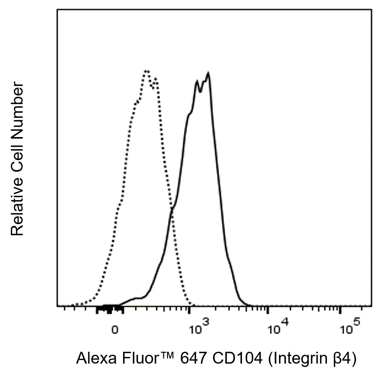
The ASC-8 monoclonal antibody specifically recognizes CD104 which is also known as Integrin beta 4 (Integrin β4 or β4 Integrin). Integrins mediate important intercellular and cell-extracellular matrix interactions which are essential for cellular adhesion and migration as well as for regulating cellular growth and survival. CD104 (Integrin β4) is an ~205 kDa single-pass type I transmembrane glycoprotein that is encoded by ITGB4 (Integrin subunit beta 4). CD104 (Integrin β4) noncovalently associates with CD49f (Integrin α6 chain) to form the Integrin α6/β4 (CD49f/CD104) complex. CD104 (Integrin β4) is expressed on basal epithelial cells, Schwann cells, and some endothelial cells and tumor cells. The CD49f/CD104 complex serves as an adhesion receptor that binds to laminins and keratin filaments. It is a component of epidermal hemidesmosomes that facilitate the stable adhesion of basal epithelial cells to the underlying basement membrane.
Development References (10)
-
Egles C, Huet HA, Dogan F, et al. Integrin-blocking antibodies delay keratinocyte re-epithelialization in a human three-dimensional wound healing model.. PLoS One. 2010; 5(5):e10528. (Clone-specific: Flow cytometry, Functional assay). View Reference
-
Hemler ME, Crouse C, Sonnenberg A. Association of the VLA alpha 6 subunit with a novel protein. A possible alternative to the common VLA beta 1 subunit on certain cell lines. J Biol Chem. 1989; 264(11):6529-6535. (Biology). View Reference
-
Jones JC, Kurpakus MA, Cooper HM, Quaranta V. A function for the integrin alpha 6 beta 4 in the hemidesmosome. Cell Regul. 1991; 2(6):427-438. (Biology). View Reference
-
Kennel SJ, Epler RG, Lankford TK, et al. Second generation monoclonal antibodies to the human integrin alpha 6 beta 4. Hybridoma. 1990; 9(3):243-255. (Biology). View Reference
-
Mohri T, Adachi Y, Ikehara S, Hioki K, Tokunaga R, Taketani S. Activated Rac1 selectively up-regulates the expression of integrin alpha6beta4 and induces cell adhesion and membrane ruffles of nonadherent colon cancer Colo201 cells.. Exp Cell Res. 1999; 253(2):533-40. (Clone-specific: Flow cytometry, Functional assay). View Reference
-
Schlossman SF. Stuart F. Schlossman .. et al., ed. Leucocyte typing V : white cell differentiation antigens : proceedings of the fifth international workshop and conference held in Boston, USA, 3-7 November, 1993. Oxford: Oxford University Press; 1995.
-
Skubitz AP, Bast RC, Wayner EA, Letourneau PC, Wilke MS. Expression of alpha 6 and beta 4 integrins in serous ovarian carcinoma correlates with expression of the basement membrane protein laminin.. Am J Pathol. 1996; 148(5):1445-61. (Immunogen: Flow cytometry). View Reference
-
Sonnenberg A, Calafat J, Janssen H, et al. Integrin alpha 6/beta 4 complex is located in hemidesmosomes, suggesting a major role in epidermal cell-basement membrane adhesion.. J Cell Biol. 1991; 113(4):907-17. (Biology). View Reference
-
Tamura RN, Rozzo C, Starr L, et al. Epithelial integrin alpha 6 beta 4: complete primary structure of alpha 6 and variant forms of beta 4.. J Cell Biol. 1990; 111(4):1593-604. (Biology). View Reference
-
Tanaka Y, Adachi T, Gianocotti F. CD104 Workshop Panel report. In: Kishimoto T. Tadamitsu Kishimoto .. et al., ed. Leucocyte typing VI : white cell differentiation antigens : proceedings of the sixth international workshop and conference held in Kobe, Japan, 10-14 November 1996. New York: Garland Pub.; 1997:432-434.
Please refer to Support Documents for Quality Certificates
Global - Refer to manufacturer's instructions for use and related User Manuals and Technical data sheets before using this products as described
Comparisons, where applicable, are made against older BD Technology, manual methods or are general performance claims. Comparisons are not made against non-BD technologies, unless otherwise noted.
For Research Use Only. Not for use in diagnostic or therapeutic procedures.