Dear Customer, If you experience any text readability or display issues on our website, please update your web browser to the latest version
.
See More
Dear Customer, If you experience any text readability or display issues on our website, please update your web browser to the latest version
.
See MoreDear Customer, If you experience any text readability or display issues on our website, please update your web browser to the latest version
.
This page has been recently translated and is available in French now.
Looks like you're visiting us from {countryName}.
Would you like to stay on the current location site or be switched to your location?
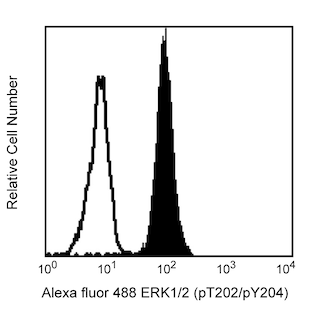
Analysis of eIF4E (pS209) in monocytes. Human peripheral blood mononuclear cells (PBMC) were either stimulated with 40 nM Phorbol 12-myristate 13-acetate (PMA, Sigma-Aldrich Cat. No. P8139) for 15 minutes (shaded histogram) or unstimulated (open histogram). The cells were fixed (BD Cytofix™ buffer, Cat. No. 554655) for 10 minutes, then permeabilized (BD Phosflow™ Perm Buffer II, Cat. No. 558052) on ice for at least 30 minutes, and then stained with Alexa Fluor® 647 Mouse anti-elF4E (pS209, Cat. No. 560240). Monocytes were selected by scatter profile. Flow cytometry was performed on a BD FACSArray™ bioanalyzer system.
The specificity of Alexa Fluor® 647 Mouse anti-elF4E(pS209) was confirmed by western blot analysis. Lysates from control populations (left blot) and PMA-treated populations (right blot) were treated with unconjugated antibody. ElF4E (pS209) is identified as a band of 25 kDa, with increased intensity in the treated cells.
Analysis of eIF4E (pS209) in monocytes. Human peripheral blood mononuclear cells (PBMC) were either stimulated with 40 nM Phorbol 12-myristate 13-acetate (PMA, Sigma-Aldrich Cat. No. P8139) for 15 minutes (shaded histogram) or unstimulated (open histogram). The cells were fixed (BD Cytofix™ buffer, Cat. No. 554655) for 10 minutes, then permeabilized (BD Phosflow™ Perm Buffer II, Cat. No. 558052) on ice for at least 30 minutes, and then stained with Alexa Fluor® 647 Mouse anti-elF4E (pS209, Cat. No. 560240). Monocytes were selected by scatter profile. Flow cytometry was performed on a BD FACSArray™ bioanalyzer system.
The specificity of Alexa Fluor® 647 Mouse anti-elF4E(pS209) was confirmed by western blot analysis. Lysates from control populations (left blot) and PMA-treated populations (right blot) were treated with unconjugated antibody. ElF4E (pS209) is identified as a band of 25 kDa, with increased intensity in the treated cells.
Analysis of eIF4E (pS209) in monocytes. Human peripheral blood mononuclear cells (PBMC) were either stimulated with 40 nM Phorbol 12-myristate 13-acetate (PMA, Sigma-Aldrich Cat. No. P8139) for 15 minutes (shaded histogram) or unstimulated (open histogram). The cells were fixed (BD Cytofix™ buffer, Cat. No. 554655) for 10 minutes, then permeabilized (BD Phosflow™ Perm Buffer II, Cat. No. 558052) on ice for at least 30 minutes, and then stained with Alexa Fluor® 647 Mouse anti-elF4E (pS209, Cat. No. 560240). Monocytes were selected by scatter profile. Flow cytometry was performed on a BD FACSArray™ bioanalyzer system.
The specificity of Alexa Fluor® 647 Mouse anti-elF4E(pS209) was confirmed by western blot analysis. Lysates from control populations (left blot) and PMA-treated populations (right blot) were treated with unconjugated antibody. ElF4E (pS209) is identified as a band of 25 kDa, with increased intensity in the treated cells.
Any use of products other than the permitted use without the express written authorization of Becton, Dickinson and Company is strictly prohibited.
Either BD Cytofix™ fixation buffer or BD Phosflow™ Fix Buffer I may be used for cell fixation. Any of the three BD Phosflow™ permeabilization buffers may be used.
The eukaryotic translation Initiation Factor 4E (eIF4E) is a 25-kDa phosphoprotein that specifically binds to the 7-methylguanosine-containing cap of mRNA. eIF4E is the rate-limiting component for the initiation of cap-dependent translation by the eIF4F translation initiation complex, which is composed of eIF4E, eIF4G, and eIF4A. This complex promotes the unwinding of secondary structure at the 5' untranslated region of mRNA that is necessary to expose and locate the AUG initiation codon. Other functions of eIF4E have been identified, such as promoting the export of mRNAs that are involved in cell cycle progression from the nucleus and differentially regulating the translation of certain mRNAs in the cytoplasm. Three mechanisms for eIF4E regulation have been identified: Mnk1-mediated phosphorylation on serine 209 (S209) is required for eIF4E binding to the cap structure; over-expression of phosphorylated eIF4E can lead to increased cell proliferation, suppression of apoptosis, and a transformed phenotype; and interactions with nonphosphorylated eIF4E-binding proteins inhibit the formation of the eIF4F complex.
The J77-925 monoclonal antibody recognizes the phosphorylated S209 (pS209) of eIF4E.
Please refer to Support Documents for Quality Certificates
Global - Refer to manufacturer's instructions for use and related User Manuals and Technical data sheets before using this products as described
Comparisons, where applicable, are made against older BD Technology, manual methods or are general performance claims. Comparisons are not made against non-BD technologies, unless otherwise noted.
Please refer to Support Documents for Quality Certificates
Global - Refer to manufacturer's instructions for use and related User Manuals and Technical data sheets before using this products as described
Comparisons, where applicable, are made against older BD Technology, manual methods or are general performance claims. Comparisons are not made against non-BD technologies, unless otherwise noted.
For Research Use Only. Not for use in diagnostic or therapeutic procedures.