-
Reagents
- Flow Cytometry Reagents
-
Western Blotting and Molecular Reagents
- Immunoassay Reagents
-
Single-Cell Multiomics Reagents
- BD® OMICS-Guard Sample Preservation Buffer
- BD® AbSeq Assay
- BD® Single-Cell Multiplexing Kit
- BD Rhapsody™ ATAC-Seq Assays
- BD Rhapsody™ Whole Transcriptome Analysis (WTA) Amplification Kit
- BD Rhapsody™ TCR/BCR Next Multiomic Assays
- BD Rhapsody™ Targeted mRNA Kits
- BD Rhapsody™ Accessory Kits
- BD® OMICS-One Protein Panels
- BD OMICS-One™ WTA Next Assay
-
Functional Assays
-
Microscopy and Imaging Reagents
-
Cell Preparation and Separation Reagents
Old Browser
This page has been recently translated and is available in French now.
Looks like you're visiting us from {countryName}.
Would you like to stay on the current location site or be switched to your location?
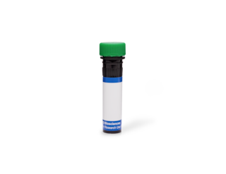
BD Transduction Laboratories™ Purified Mouse Anti-ERK (pan ERK)
Clone 16/ERK (pan ERK) (RUO)


Western blot analysis of ERK (pan ERK) on a RSV-3T3 cell lysate. Lane 1: 1:5000, lane 2: 1:10,000, lane 3: 1:20,000 dilution of the mouse anti-ERK (pan ERK) antibody.
Immunohistochemical staining on a rat brain formalin-fixed paraffin-embedded section with citrate buffer pretreatment (20X magnification).

Western blot analysis of ERK (pan ERK) on a RSV-3T3 cell lysate. Lane 1: 1:5000, lane 2: 1:10,000, lane 3: 1:20,000 dilution of the mouse anti-ERK (pan ERK) antibody.

Immunohistochemical staining on a rat brain formalin-fixed paraffin-embedded section with citrate buffer pretreatment (20X magnification).

Immunofluorescence staining of 3T3-L1 cells (Mouse embryonic fibroblasts; ATCC CL-173).


Regulatory Status Legend
Any use of products other than the permitted use without the express written authorization of Becton, Dickinson and Company is strictly prohibited.
Preparation And Storage
Recommended Assay Procedures
Western blot: Please refer to http://www.bdbiosciences.com/pharmingen/protocols/Western_Blotting.shtml
Product Notices
- Since applications vary, each investigator should titrate the reagent to obtain optimal results.
- Please refer to www.bdbiosciences.com/us/s/resources for technical protocols.
- Caution: Sodium azide yields highly toxic hydrazoic acid under acidic conditions. Dilute azide compounds in running water before discarding to avoid accumulation of potentially explosive deposits in plumbing.
- Source of all serum proteins is from USDA inspected abattoirs located in the United States.
Companion Products


The family of serine/threonine kinases known as ERKs (extracellular signal regulated kinases) or MAPKs (mitogen-activated protein kinases) are activated after cell stimulation by a variety of hormones and growth factors. Cell stimulation induces a signaling cascade that leads to phosphorylation of MEK (MAPK/ERK kinase) which, in turn, activates ERK via tyrosine and threonine phosphorylation. A myriad of proteins represent the downstream effectors for the active ERK and implicate it in the control of cell proliferation and differentiation, as well as regulation of the cytoskeleton. Activation of ERK is normally transient and cells possess dual specificity phosphatases that are responsible for its down-regulation. Furthermore, multiple studies have shown that elevated ERK activity is associated with some cancers. ERK1 may be observable migrating at 44 kDa and ERK2 at 42 kDa in addition to a 54 kDa ERK and a MAP kinase in the 90 kDa range.
Development References (5)
-
Jiang K, Zhong B, Gilvary DL, et al. Syk regulation of phosphoinositide 3-kinase-dependent NK cell function. J Immunol. 2002; 168(7):3155-3164. (Biology: Western blot). View Reference
-
Maher P. Phorbol esters inhibit fibroblast growth factor-2-stimulated fibroblast proliferation by a p38 MAP kinase dependent pathway. Oncogene. 2002; 21(13):1978-1988. (Biology: Western blot). View Reference
-
Nutt SL, Dingwell KS, Holt CE, Amaya E. Xenopus Sprouty2 inhibits FGF-mediated gastrulation movements but does not affect mesoderm induction and patterning. Genes Dev. 2001; 15(9):1152-1166. (Biology: Western blot). View Reference
-
Reszka AA, Seger R, Diltz CD, Krebs EG, Fischer EH. Association of mitogen-activated protein kinase with the microtubule cytoskeleton. Proc Natl Acad Sci U S A. 1995; 92(19):8881-8885. (Biology: Immunofluorescence, Western blot). View Reference
-
Watson FL, Heerssen HM, Bhattacharyya A, Klesse L, Lin MZ, Segal RA. Neurotrophins use the Erk5 pathway to mediate a retrograde survival response. Nat Neurosci. 2001; 4(10):981-988. (Biology: Immunoprecipitation). View Reference
Please refer to Support Documents for Quality Certificates
Global - Refer to manufacturer's instructions for use and related User Manuals and Technical data sheets before using this products as described
Comparisons, where applicable, are made against older BD Technology, manual methods or are general performance claims. Comparisons are not made against non-BD technologies, unless otherwise noted.
For Research Use Only. Not for use in diagnostic or therapeutic procedures.