-
Reagents
- Flow Cytometry Reagents
-
Western Blotting and Molecular Reagents
- Immunoassay Reagents
-
Single-Cell Multiomics Reagents
- BD OMICS-Guard™ Sample Preservation Buffer
- BD® AbSeq Assay
- BD® Single-Cell Multiplexing Kit
- BD Rhapsody™ ATAC-Seq Assays
- BD Rhapsody™ Whole Transcriptome Analysis (WTA) Amplification Kit
- BD Rhapsody™ TCR/BCR Next Multiomic Assays
- BD Rhapsody™ Targeted mRNA Kits
- BD Rhapsody™ Accessory Kits
- BD® OMICS-One Protein Panels
- BD OMICS-One™ WTA Next Assay
-
Functional Assays
-
Microscopy and Imaging Reagents
-
Cell Preparation and Separation Reagents
Old Browser
This page has been recently translated and is available in French now.
Looks like you're visiting us from {countryName}.
Would you like to stay on the current location site or be switched to your location?
BD Phosflow™ Alexa Fluor® 647 Mouse Anti-ZAP70 (PY319)/Syk (PY352)
Clone 17A/P-ZAP70 (RUO)
Flow cytometric analysis of ZAP70 (Y319)/Syk (Y352). Jurkat cells (Human T-cell leukemia; ATCC TIB-152) were starved overnight in RPMI containing 0.1% FCS. The following day, cells were either left untreated (unshaded) or treated (shaded) with H2O2 (5 mM for 15 minutes at 37°C). Cells were fixed in BD Cytofix™ buffer for 10 minutes at 37°C and permeabilized with BD Phosflow™ Perm Buffer III for 30 minutes on ice. Cells were then stained with the Alexa Fluor® 647 Zap70 (Y319)/Syk (Y352) antibody. The cells were analyzed on a BD FACSCalibur™ flow cytometry instrument.
Regulatory Status Legend
Any use of products other than the permitted use without the express written authorization of Becton, Dickinson and Company is strictly prohibited.
Preparation And Storage
Recommended Assay Procedures
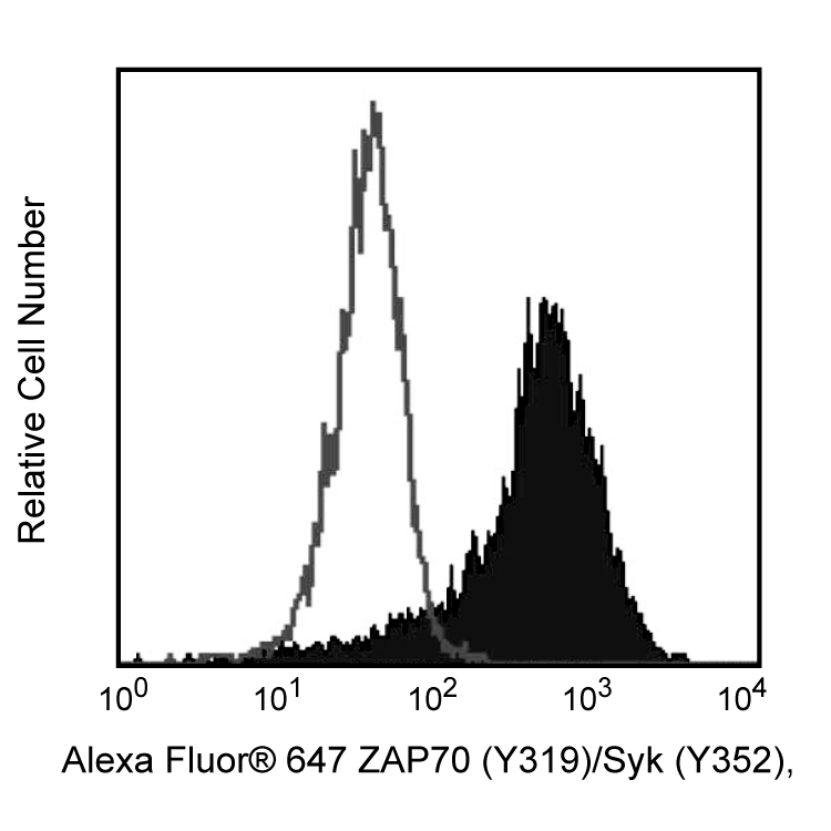
Jurkat cells treated with H2O2 are suggested as a positive control. However, other cell types or methods may also be used for detection of phosphorylated ZAP70.
BD® CompBeads can be used as surrogates to assess fluorescence spillover (compensation). When fluorochrome conjugated antibodies are bound to BD® CompBeads, they have spectral properties very similar to cells. However, for some fluorochromes there can be small differences in spectral emissions compared to cells, resulting in spillover values that differ when compared to biological controls. It is strongly recommended that when using a reagent for the first time, users compare the spillover on cell and BD® CompBeads to ensure that BD® CompBeads are appropriate for your specific cellular application.
Product Notices
- This reagent has been pre-diluted for use at the recommended Volume per Test. We typically use 1 × 10^6 cells in a 100-µl experimental sample (a test).
- Alexa Fluor® 647 fluorochrome emission is collected at the same instrument settings as for allophycocyanin (APC).
- Source of all serum proteins is from USDA inspected abattoirs located in the United States.
- Caution: Sodium azide yields highly toxic hydrazoic acid under acidic conditions. Dilute azide compounds in running water before discarding to avoid accumulation of potentially explosive deposits in plumbing.
- For fluorochrome spectra and suitable instrument settings, please refer to our Multicolor Flow Cytometry web page at www.bdbiosciences.com/colors.
- Please refer to www.bdbiosciences.com/us/s/resources for technical protocols.
- This product is provided under an intellectual property license between Life Technologies Corporation and BD Businesses. The purchase of this product conveys to the buyer the non-transferable right to use the purchased amount of the product and components of the product in research conducted by the buyer (whether the buyer is an academic or for-profit entity). The buyer cannot sell or otherwise transfer (a) this product (b) its components or (c) materials made using this product or its components to a third party or otherwise use this product or its components or materials made using this product or its components for Commercial Purposes. Commercial Purposes means any activity by a party for consideration and may include, but is not limited to: (1) use of the product or its components in manufacturing; (2) use of the product or its components to provide a service, information, or data; (3) use of the product or its components for therapeutic, diagnostic or prophylactic purposes; or (4) resale of the product or its components, whether or not such product or its components are resold for use in research. For information on purchasing a license to this product for any other use, contact Life Technologies Corporation, Cell Analysis Business Unit Business Development, 29851 Willow Creek Road, Eugene, OR 97402, USA, Tel: (541) 465-8300. Fax: (541) 335-0504.
- Please refer to http://regdocs.bd.com to access safety data sheets (SDS).
- Alexa Fluor™ is a trademark of Life Technologies Corporation.
ZAP70 is a protein tyrosine kinase (PTK) that associates with the z subunit of the T cell antigen receptor (TCR) and undergoes tyrosine phosphorylation following TCR stimulation. ZAP70 contains two SH2-like domains with the PTK domain located at the C-terminus. It appears that both ZAP70 and Syk are recruited to the phosphorylated CD3 and z subunits after TCR stimulation. TCR stimulation leads to autophosphorylation of ZAP70 at Tyr-315 amd Tyr-319, and mutation of the Tyr-319 site dramatically impairs TCR signaling. In addition, TCR-mediated Lck activity leads to phosphorylation of ZAP70 on Tyr-493 in the regulatory loop of the kinase domain leading to upregulation of ZAP70 kinase activity. The significance of ZAP70 activation in mediating TCR signal transduction has been confirmed by showing that ZAP70 activity is absent in an autosomal recessive form of severe combined immunodeficiency (SCID). This is due to mutations affecting the ZAP70 kinase domain which affect the stability of the protein and TCR signaling.
Clone 17A/P-ZAP70 recognizes the phosphorylated form of ZAP70 (Y319). It also cross-reacts with SYK (Y352) due to homology of the phosphorylation site with ZAP70 (Y319). The PE-conjugated format has been evaluated using human and mouse model systems. The unconjugated form of the antibody (Cat. No. 612574) has also been shown to work in western blot analysis on human, mouse, and rat cells.
Development References (3)
-
Arpaia E, Shahar M, Dadi H, Cohen A, Roifman CM. Defective T cell receptor signaling and CD8+ thymic selection in humans lacking zap-70 kinase. Cell. 1994; 76(5):947-958. (Biology). View Reference
-
Chan AC, Kadlecek TA, Elder ME, et al. ZAP-70 deficiency in an autosomal recessive form of severe combined immunodeficiency. Science. 1994; 264:1599-1601. (Biology).
-
Di Bartolo V, Mege D, Germain V, et al. Tyrosine 319, a newly identified phosphorylation site of ZAP-70, plays a critical role in T cell antigen receptor signaling. J Biol Chem. 1999; 274(10):6285-6294. (Biology). View Reference
Please refer to Support Documents for Quality Certificates
Global - Refer to manufacturer's instructions for use and related User Manuals and Technical data sheets before using this products as described
Comparisons, where applicable, are made against older BD Technology, manual methods or are general performance claims. Comparisons are not made against non-BD technologies, unless otherwise noted.
For Research Use Only. Not for use in diagnostic or therapeutic procedures.