The site is undergoing maintenance on
Thursday, September 10, 5:00 pm through Friday, September 11, 1:00 am (CEST), 2026.
If you encounter problems while ordering, please contact us at BDBCustomerService@bd.com
See More
The site is undergoing maintenance on
Thursday, September 10, 5:00 pm through Friday, September 11, 1:00 am (CEST), 2026.
If you encounter problems while ordering, please contact us at BDBCustomerService@bd.com
See MoreThe site is undergoing maintenance on
Thursday, September 10, 5:00 pm through Friday, September 11, 1:00 am (CEST), 2026
.If you encounter problems while ordering, please contact us at BDBCustomerService@bd.com
This page has been recently translated and is available in French now.
Looks like you're visiting us from {countryName}.
Would you like to stay on the current location site or be switched to your location?
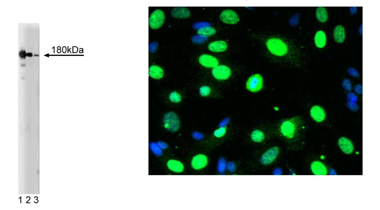
Western blot analysis of NMDAR2B on a rat cerebrum lysate (left). Lane 1: 1:250, lane 2: 1:500, lane 3: 1:1000 dilution of the mouse anti- NMDAR2B antibody.
Immunofluorescent staining of SK-N-SH cells (right). Cells were seeded in a 384 well collagen coated Microplates (Material # 353962) at ~ 8,000 cells per well. After overnight incubation, cells were stained using the Triton X100 fix/perm protocol (see Recommended Assay Procedure; Bioimaging protocol link) and the anti- NMDAR2B antibody. The second step reagent was Alexa Fluor® 488 goat anti mouse Ig (Invitrogen)(pseudo colored green). Cell nuclei were counter stained with Hoechst 33342 (pseudo colored blue). The image was taken on a BD Pathway™ 855 or 435 Bioimager System using a 20x objective and merged using the BD AttoVison ™ software. This antibody also stained SH-SY5Y, SK-N-SH, C6, U87 and U373 cells using both the Triton X100 and methanol fix/perm protocols (see Recommended Assay Procedure; Bioimaging protocol link).
Western blot analysis of NMDAR2B on a rat cerebrum lysate (left). Lane 1: 1:250, lane 2: 1:500, lane 3: 1:1000 dilution of the mouse anti- NMDAR2B antibody.
Immunofluorescent staining of SK-N-SH cells (right). Cells were seeded in a 384 well collagen coated Microplates (Material # 353962) at ~ 8,000 cells per well. After overnight incubation, cells were stained using the Triton X100 fix/perm protocol (see Recommended Assay Procedure; Bioimaging protocol link) and the anti- NMDAR2B antibody. The second step reagent was Alexa Fluor® 488 goat anti mouse Ig (Invitrogen)(pseudo colored green). Cell nuclei were counter stained with Hoechst 33342 (pseudo colored blue). The image was taken on a BD Pathway™ 855 or 435 Bioimager System using a 20x objective and merged using the BD AttoVison ™ software. This antibody also stained SH-SY5Y, SK-N-SH, C6, U87 and U373 cells using both the Triton X100 and methanol fix/perm protocols (see Recommended Assay Procedure; Bioimaging protocol link).
Western blot analysis of NMDAR2B on a rat cerebrum lysate (left). Lane 1: 1:250, lane 2: 1:500, lane 3: 1:1000 dilution of the mouse anti- NMDAR2B antibody.
Immunofluorescent staining of SK-N-SH cells (right). Cells were seeded in a 384 well collagen coated Microplates (Material # 353962) at ~ 8,000 cells per well. After overnight incubation, cells were stained using the Triton X100 fix/perm protocol (see Recommended Assay Procedure; Bioimaging protocol link) and the anti- NMDAR2B antibody. The second step reagent was Alexa Fluor® 488 goat anti mouse Ig (Invitrogen)(pseudo colored green). Cell nuclei were counter stained with Hoechst 33342 (pseudo colored blue). The image was taken on a BD Pathway™ 855 or 435 Bioimager System using a 20x objective and merged using the BD AttoVison ™ software. This antibody also stained SH-SY5Y, SK-N-SH, C6, U87 and U373 cells using both the Triton X100 and methanol fix/perm protocols (see Recommended Assay Procedure; Bioimaging protocol link).
Western blot analysis of NMDAR2B on a rat cerebrum lysate (left). Lane 1: 1:250, lane 2: 1:500, lane 3: 1:1000 dilution of the mouse anti- NMDAR2B antibody.
Immunofluorescent staining of SK-N-SH cells (right). Cells were seeded in a 384 well collagen coated Microplates (Material # 353962) at ~ 8,000 cells per well. After overnight incubation, cells were stained using the Triton X100 fix/perm protocol (see Recommended Assay Procedure; Bioimaging protocol link) and the anti- NMDAR2B antibody. The second step reagent was Alexa Fluor® 488 goat anti mouse Ig (Invitrogen)(pseudo colored green). Cell nuclei were counter stained with Hoechst 33342 (pseudo colored blue). The image was taken on a BD Pathway™ 855 or 435 Bioimager System using a 20x objective and merged using the BD AttoVison ™ software. This antibody also stained SH-SY5Y, SK-N-SH, C6, U87 and U373 cells using both the Triton X100 and methanol fix/perm protocols (see Recommended Assay Procedure; Bioimaging protocol link).

Western blot analysis of NMDAR2B on a rat cerebrum lysate (left). Lane 1: 1:250, lane 2: 1:500, lane 3: 1:1000 dilution of the mouse anti- NMDAR2B antibody.
Immunofluorescent staining of SK-N-SH cells (right). Cells were seeded in a 384 well collagen coated Microplates (Material # 353962) at ~ 8,000 cells per well. After overnight incubation, cells were stained using the Triton X100 fix/perm protocol (see Recommended Assay Procedure; Bioimaging protocol link) and the anti- NMDAR2B antibody. The second step reagent was Alexa Fluor® 488 goat anti mouse Ig (Invitrogen)(pseudo colored green). Cell nuclei were counter stained with Hoechst 33342 (pseudo colored blue). The image was taken on a BD Pathway™ 855 or 435 Bioimager System using a 20x objective and merged using the BD AttoVison ™ software. This antibody also stained SH-SY5Y, SK-N-SH, C6, U87 and U373 cells using both the Triton X100 and methanol fix/perm protocols (see Recommended Assay Procedure; Bioimaging protocol link).
Western blot analysis of NMDAR2B on a rat cerebrum lysate (left). Lane 1: 1:250, lane 2: 1:500, lane 3: 1:1000 dilution of the mouse anti- NMDAR2B antibody.
Immunofluorescent staining of SK-N-SH cells (right). Cells were seeded in a 384 well collagen coated Microplates (Material # 353962) at ~ 8,000 cells per well. After overnight incubation, cells were stained using the Triton X100 fix/perm protocol (see Recommended Assay Procedure; Bioimaging protocol link) and the anti- NMDAR2B antibody. The second step reagent was Alexa Fluor® 488 goat anti mouse Ig (Invitrogen)(pseudo colored green). Cell nuclei were counter stained with Hoechst 33342 (pseudo colored blue). The image was taken on a BD Pathway™ 855 or 435 Bioimager System using a 20x objective and merged using the BD AttoVison ™ software. This antibody also stained SH-SY5Y, SK-N-SH, C6, U87 and U373 cells using both the Triton X100 and methanol fix/perm protocols (see Recommended Assay Procedure; Bioimaging protocol link).
Any use of products other than the permitted use without the express written authorization of Becton, Dickinson and Company is strictly prohibited.
The majority of synapses in the central nervous system utilize glutamate as a neurotransmitter to produce rapid neuronal excitation. Glutamate has a diverse array of receptors that can be categorized into two groups: ionotropic and metabotropic. The ionotropic receptors are subdivided into two distint types: 1) receptors for N-methyl D-aspartate (NMDAR) and 2) non-NMDA receptors for AMPA and kainate. Three types of NMDAR2 have been identified: NR2A, NR2B, and NR2C. NR2A and NR2B contain a C-terminal extension (>600 amino acids) that has small scattered regions of conserved sequence. The three NR2 mRNAs show overlapping, differential expression patterns in the rat brain. NR2B has been reported to be expressed in the forebrain, thalamic nuclei, amygdaloid nuclei, caudateputamen, and in restricted regions of the olfactory bulb.
Please refer to Support Documents for Quality Certificates
Global - Refer to manufacturer's instructions for use and related User Manuals and Technical data sheets before using this products as described
Comparisons, where applicable, are made against older BD Technology, manual methods or are general performance claims. Comparisons are not made against non-BD technologies, unless otherwise noted.
For Research Use Only. Not for use in diagnostic or therapeutic procedures.