Old Browser
This page has been recently translated and is available in French now.
Looks like you're visiting us from {countryName}.
Would you like to stay on the current country site or be switched to your country?


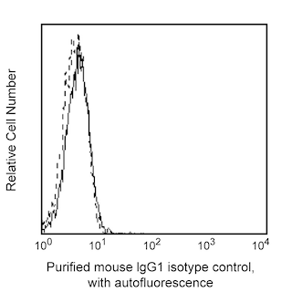
Flow cytometric analysis of CD28 expression on mouse splenocytes. BALB/c mouse splenic leucocytes were preincubated with Purified Rat Anti-Mouse CD16/CD32 antibody (Mouse BD Fc Block™) (Cat. No. 553141/553142). The cells were then stained with either Purified Mouse IgG1, κ Isotype Control (Cat. No. 556648; dashed line histogram) or Purified NA/LE Mouse Anti-Mouse CD28 antibody (Cat. No. 566883; solid line histogram) at 1 µg/test followed by Biotin Rat Anti-Mouse IgG1 antibody (Cat. No. 553441) and then PE Streptavidin (Cat. No. 554061). The fluorescence histogram showing CD28 expression (or Ig isotype control staining) was derived from gated events with the forward and side light-scatter characteristics of viable splenocytes. Flow cytometry and data analysis were performed using a BD LSRFortessa™ X-20 Analyzer System and FlowJo™ software. Data shown on this Technical Data Sheet are not lot specific.

Flow cytometric analysis of proliferating mouse splenocytes stimulated with either Anti-CD3 plus Anti-CD28 antibodies or Anti-CD28 antibody alone. C57BL/6 mouse spleen cells were treated with BD Pharm Lyse™ Lysing Buffer (Cat. No. 555899) to lyse erythrocytes. The splenic leucocytes were washed and labeled with BD Horizon™ CFSE (5 μM; Cat. No. 565082) for 10 minutes at 37°C. The cells were washed twice and then cultured with either Purified NA/LE Hamster Anti-Mouse CD3e (10 µg/ml plate-bound; Cat. No. 553057) and Purified NA/LE Mouse Anti-Mouse CD28 (1 µg/ml soluble; Cat. No. 566883) antibodies (Left Plot) or with Purified NA/LE Mouse Anti-Mouse CD28 antibody (1 μg/ml soluble) alone (Right Plot). After 3 days, the cells were harvested, washed, and stained with BD Horizon™ BUV395 Rat Anti-Mouse CD4 antibody (Cat. No. 563790). BD Pharmingen™ DAPI Solution (Cat. No. 564907) was added to samples prior to acquisition to exclude non-viable cells. Two-color flow cytometric dot plots showing the correlated expression of CFSE staining versus CD4 levels were derived from gated events with the forward and side light-scatter characteristics of viable (DAPI-negative) lymphocytes. Flow cytometry and data analysis were performed using a BD LSRFortessa™ X-20 Analyzer System and FlowJo™ software. Data shown on this Technical Data Sheet are not lot specific.


BD Pharmingen™ Purified NA/LE Mouse Anti-Mouse CD28

BD Pharmingen™ Purified NA/LE Mouse Anti-Mouse CD28

Regulatory Status Legend
Any use of products other than the permitted use without the express written authorization of Becton, Dickinson and Company is strictly prohibited.
Preparation And Storage
Product Notices
- Please refer to www.bdbiosciences.com/us/s/resources for technical protocols.
- Since applications vary, each investigator should titrate the reagent to obtain optimal results.
- Please refer to http://regdocs.bd.com to access safety data sheets (SDS).
Companion Products


The D665 monoclonal antibody specifically recognizes CD28. CD28 is a single-pass type I transmembrane glycoprotein that is encoded by Cd28 (CD28 antigen) which belongs to the immunoglobulin gene superfamily. CD28 has an immunoglobulin V-set (IgV-like) domain in its extracellular region that is followed by a transmembrane region and a cytoplasmic domain and can be expressed on the cell surface as a disulfide-linked homodimer. CD28 functions as a costimulatory receptor involved in the induction of T cell activation, proliferation and cytokine production as well as the promotion of T-cell survival. CD28 binds to B7-1/CD80 or B7-2/CD86 that are expressed on antigen-presenting cells. CD28 is expressed on thymocytes, most peripheral CD4+ T cells and CD8+ T cells, NK cells, plasmacytoid dendritic cells, and plasma cells. The D665 antibody functions as a superagonistic antibody that can stimulate T-cell proliferation in the apparent absence of antigen-mediated stimulation through the T cell receptor (TCR) for antigen. The D665 antibody reportedly binds to the C'D loop of mouse CD28, whereas the E18 antibody binds to the ligand-binding region of mouse CD28.
Development References (4)
-
Chen J, Xie L, Toyama S, et al. The effects of Foxp3-expressing regulatory T cells expanded with CD28 superagonist antibody in DSS-induced mice colitis.. Int Immunopharmacol. 2011; 11(5):610-7. (Clone-specific: In vivo exacerbation). View Reference
-
Dennehy KM, Elias F, Zeder-Lutz G, et al. Cutting edge: monovalency of CD28 maintains the antigen dependence of T cell costimulatory responses.. J Immunol. 2006; 176(10):5725-9. (Immunogen: Flow cytometry, Functional assay). View Reference
-
Gogishvili T, Langenhorst D, Lühder F, et al. Rapid regulatory T-cell response prevents cytokine storm in CD28 superagonist treated mice. PLoS One. 2009; 4(2):e4643. (Clone-specific: In vivo exacerbation). View Reference
-
Langenhorst D, Tabares P, Gulde T, et al. Self-Recognition Sensitizes Mouse and Human Regulatory T Cells to Low-Dose CD28 Superagonist Stimulation.. Front Immunol. 2017; 8:1985. (Clone-specific: In vivo exacerbation). View Reference
Please refer to Support Documents for Quality Certificates
Global - Refer to manufacturer's instructions for use and related User Manuals and Technical data sheets before using this products as described
Comparisons, where applicable, are made against older BD Technology, manual methods or are general performance claims. Comparisons are not made against non-BD technologies, unless otherwise noted.
For Research Use Only. Not for use in diagnostic or therapeutic procedures.
Report a Site Issue
This form is intended to help us improve our website experience. For other support, please visit our Contact Us page.