





.png)


Preparation And Storage


Product Notices
- This reagent has been pre-diluted for use at the recommended Volume per Test. We typically use 1 × 10^6 cells in a 100-µl experimental sample (a test).
- Please refer to www.bdbiosciences.com/us/s/resources for technical protocols.
- An isotype control should be used at the same concentration as the antibody of interest.
- For fluorochrome spectra and suitable instrument settings, please refer to our Multicolor Flow Cytometry web page at www.bdbiosciences.com/colors.
- Caution: Sodium azide yields highly toxic hydrazoic acid under acidic conditions. Dilute azide compounds in running water before discarding to avoid accumulation of potentially explosive deposits in plumbing.
- Source of all serum proteins is from USDA inspected abattoirs located in the United States.
Companion Products



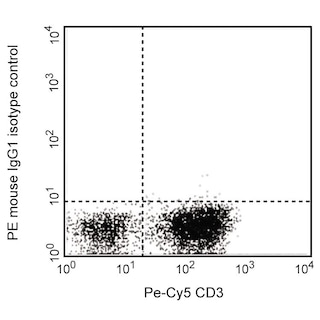
The N14-47 monoclonal antibody recognizes RNA-binding protein Musashi homolog 1 (Musashi-1 or MSI1) that represses the translation of particular mRNAs by binding specific RNA sequence motifs. MSI1 contains two RNA-recognition domains at amino acids 20-110 and 109-186. It is expressed in early lineage cells, such as neural stem/progenitor cells, small intestinal stem cells, and mammary stem cells. MSI1 has also been detected in many human cancers, including gliomas, melanomas, colorectal adenomas and adenocarcinomas, suggesting its involvement in cancer development.

Development References (3)
-
Glazer RI, Wang XY, Yuan H, Yin Y. Musashi1: a stem cell marker no longer in search of a function. Cell Cycle. 2008; 7(17):2635-2639. (Biology). View Reference
-
Montgomery RK, Breault DT. Small intestinal stem cell markers. J Anat. 2008; 213(1):52-58. (Biology). View Reference
-
Okano H, Imai T, Okabe M. Musashi: a translational regulator of cell fate. J Cell Sci. 2002; 115:1355-1359. (Biology). View Reference
Please refer to Support Documents for Quality Certificates
Global - Refer to manufacturer's instructions for use and related User Manuals and Technical data sheets before using this products as described
Comparisons, where applicable, are made against older BD Technology, manual methods or are general performance claims. Comparisons are not made against non-BD technologies, unless otherwise noted.
For Research Use Only. Not for use in diagnostic or therapeutic procedures.
