Preparation And Storage
Product Notices
- This reagent has been pre-diluted for use at the recommended Volume per Test. We typically use 1 × 10^6 cells in a 100-µl experimental sample (a test).
- An isotype control should be used at the same concentration as the antibody of interest.
- Please refer to www.bdbiosciences.com/us/s/resources for technical protocols.
- For fluorochrome spectra and suitable instrument settings, please refer to our Multicolor Flow Cytometry web page at www.bdbiosciences.com/colors.
- Source of all serum proteins is from USDA inspected abattoirs located in the United States.
- Caution: Sodium azide yields highly toxic hydrazoic acid under acidic conditions. Dilute azide compounds in running water before discarding to avoid accumulation of potentially explosive deposits in plumbing.
Companion Products


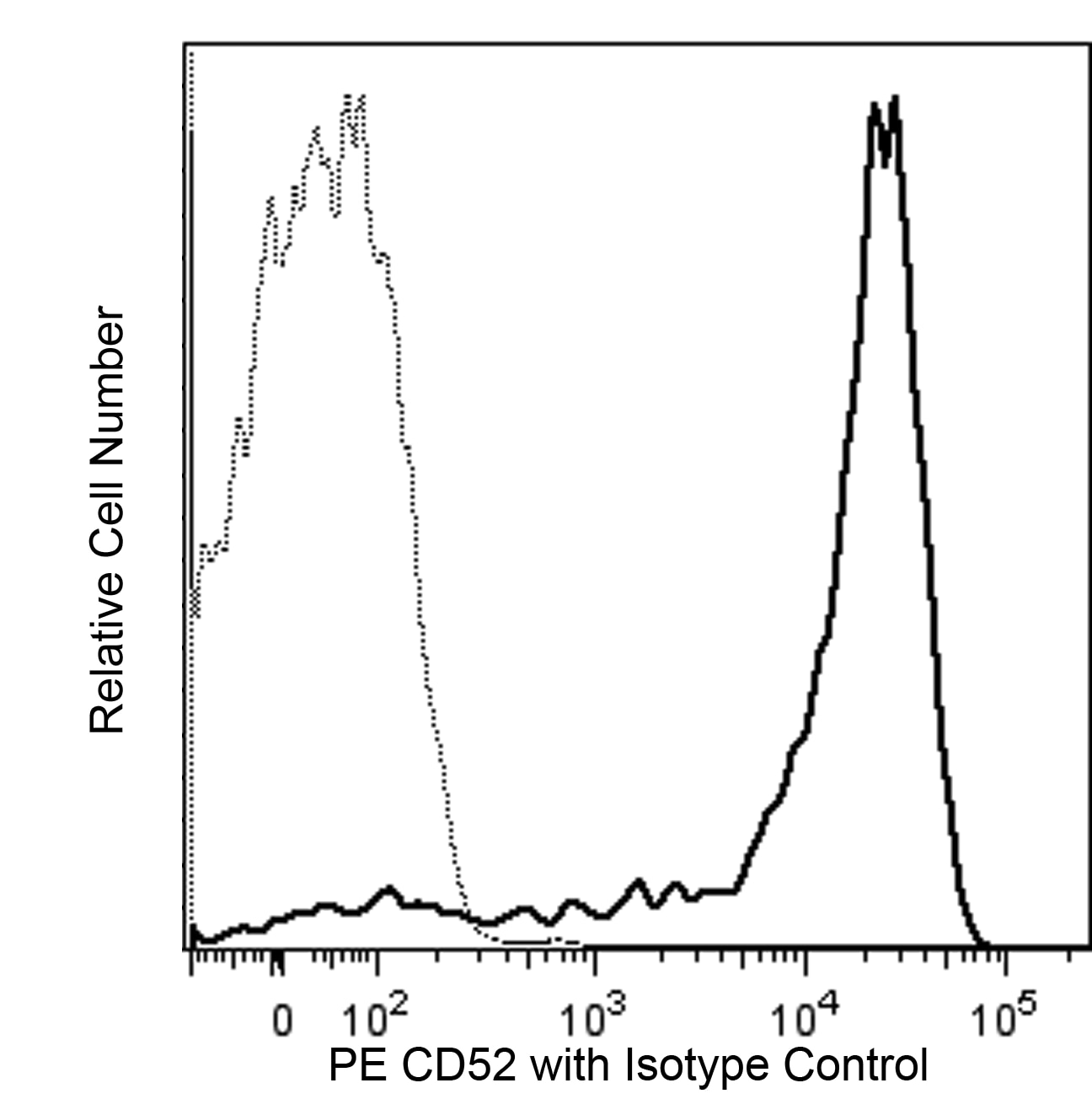
The 4C8 monoclonal antibody specifically binds to CD52 which is also known as Cambridge pathology 1 antigen (CAMPATH-1) or Human epididymis-specific protein 5 (HE5). CD52 is a highly N-glycosylated, 25-29 kDa protein whose C-terminus is glycophosphatidylinositol anchored in the membrane. It is highly expressed on the surface of thymocytes and mature lymphocytes but not on their stem cell precursors. It is also expressed on monocytes, dendritic cells, eosinophils and epithelial cells of the epididymis and seminal vesicles but not on neutrophils, plasma cells, platelets or erythrocytes. Although its functional role is not well characterized, the CD52 antigen serves as an exquisitely sensitive target antigen for antibody and complement-mediated lysis of CD52-positive cells. Anti-CD52 antibodies are being used clinically to remove lymphocytes from transplanted bone marrow cell preparations and in the treatment of some malignant diseases.

Development References (2)
-
Masuyama J, Yoshio T, Suzuki K, et al. Characterization of the 4C8 antigen involved in transendothelial migration of CD26(hi) T cells after tight adhesion to human umbilical vein endothelial cell monolayers.. J Exp Med. 1999; 189(6):979-990. (Immunogen: Blocking, Flow cytometry, Stimulation, Western blot). View Reference
-
Zola H. Leukocyte and stromal cell molecules : the CD markers. Hoboken, N.J.: Wiley-Liss; 2007.
Please refer to Support Documents for Quality Certificates
Global - Refer to manufacturer's instructions for use and related User Manuals and Technical data sheets before using this products as described
Comparisons, where applicable, are made against older BD Technology, manual methods or are general performance claims. Comparisons are not made against non-BD technologies, unless otherwise noted.
For Research Use Only. Not for use in diagnostic or therapeutic procedures.