
Preparation And Storage
Recommended Assay Procedures
BD™ CompBeads can be used as surrogates to assess fluorescence spillover (Compensation). When fluorochrome conjugated antibodies are bound to CompBeads, they have spectral properties very similar to cells. However, for some fluorochromes there can be small differences in spectral emissions compared to cells, resulting in spillover values that differ when compared to biological controls. It is strongly recommended that when using a reagent for the first time, users compare the spillover on cell and CompBead to ensure that BD Comp beads are appropriate for your specific cellular application.
Product Notices
- Since applications vary, each investigator should titrate the reagent to obtain optimal results.
- An isotype control should be used at the same concentration as the antibody of interest.
- Caution: Sodium azide yields highly toxic hydrazoic acid under acidic conditions. Dilute azide compounds in running water before discarding to avoid accumulation of potentially explosive deposits in plumbing.
- For fluorochrome spectra and suitable instrument settings, please refer to our Multicolor Flow Cytometry web page at www.bdbiosciences.com/colors.
- Please refer to http://regdocs.bd.com to access safety data sheets (SDS).
- Although hamster immunoglobulin isotypes have not been well defined, BD Biosciences Pharmingen has grouped Armenian and Syrian hamster IgG monoclonal antibodies according to their reactivity with a panel of mouse anti-hamster IgG mAbs. A table of the hamster IgG groups, Reactivity of Mouse Anti-Hamster Ig mAbs, may be viewed at http://www.bdbiosciences.com/documents/hamster_chart_11x17.pdf.
- Please refer to www.bdbiosciences.com/us/s/resources for technical protocols.
Companion Products


.png?imwidth=320)
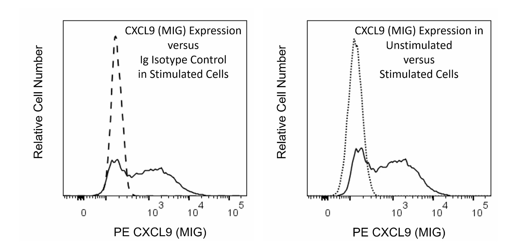
The MIG 2F5.5 monoclonal antibody specifically recognizes MIG (Monokine induced by gamma interferon) which is also known as C-X-C motif chemokine 9 (CXCL9) or Small-inducible cytokine B9 (Scyb9). Mouse CXCL9 (MIG) is encoded by Cxcl9 and belongs to the C-X-C family of chemokines. CXCL9 (MIG) production is induced in a variety of cells including macrophages, hepatocytes, and endothelial cells in response to gamma interferon (IFN-γ). The seven-transmembrane G-protein coupled receptor termed CXCR3, also known as CD183, serves as the cell surface signaling receptor for CXCL9 (MIG). This chemotactic factor attracts activated and memory CD4+ and CD8+ T cells including Type-1 (Th1-like) CD4+ T cells as well as natural killer (NK) cells. CXCL9 (MIG) plays important roles in promoting protective immunity through the attraction of CXCR3+ T cells and NK cells but can also function in inflammation and autoimmune diseases.

Development References (4)
-
Asai A, Tsuda Y, Kobayashi M, Hanafusa T, Herndon DN, Suzuki F. Pathogenic role of macrophages in intradermal infection of methicillin-resistant Staphylococcus aureus in thermally injured mice.. Infect Immun. 2010; 78(10):4311-9. (Clone-specific: Flow cytometry). View Reference
-
Krug A, Uppaluri R, Facchetti F, et al. IFN-producing cells respond to CXCR3 ligands in the presence of CXCL12 and secrete inflammatory chemokines upon activation. J Immunol. 2002; 169(11):6079-6083. (Immunogen: ELISA). View Reference
-
Marcus AJ, Ullman HL, Safier LB. Lipid composition of subcellular particles of human blood platelets.. J Lipid Res. 1969; 10(1):108-14. (Biology). View Reference
-
Sung JH, Zhang H, Moseman EA, et al. Chemokine guidance of central memory T cells is critical for antiviral recall responses in lymph nodes.. Cell. 2012; 150(6):1249-63. (Clone-specific: Flow cytometry, Immunofluorescence). View Reference
Please refer to Support Documents for Quality Certificates
Global - Refer to manufacturer's instructions for use and related User Manuals and Technical data sheets before using this products as described
Comparisons, where applicable, are made against older BD Technology, manual methods or are general performance claims. Comparisons are not made against non-BD technologies, unless otherwise noted.
For Research Use Only. Not for use in diagnostic or therapeutic procedures.