Old Browser
This page has been recently translated and is available in French now.
Looks like you're visiting us from {countryName}.
Would you like to stay on the current country site or be switched to your country?


Regulatory Status Legend
Any use of products other than the permitted use without the express written authorization of Becton, Dickinson and Company is strictly prohibited.
Preparation And Storage
Recommended Assay Procedures
For optimal and reproducible results, BD Horizon Brilliant Stain Buffer should be used anytime two or more BD Horizon Brilliant dyes (including BD OptiBuild Brilliant reagents) are used in the same experiment. Fluorescent dye interactions may cause staining artifacts which may affect data interpretation. The BD Horizon Brilliant Stain Buffer was designed to minimize these interactions. More information can be found in the Technical Data Sheet of the BD Horizon Brilliant Stain Buffer (Cat. No. 563794).
Product Notices
- This antibody was developed for use in flow cytometry.
- The production process underwent stringent testing and validation to assure that it generates a high-quality conjugate with consistent performance and specific binding activity. However, verification testing has not been performed on all conjugate lots.
- Researchers should determine the optimal concentration of this reagent for their individual applications.
- An isotype control should be used at the same concentration as the antibody of interest.
- Caution: Sodium azide yields highly toxic hydrazoic acid under acidic conditions. Dilute azide compounds in running water before discarding to avoid accumulation of potentially explosive deposits in plumbing.
- For fluorochrome spectra and suitable instrument settings, please refer to our Multicolor Flow Cytometry web page at www.bdbiosciences.com/colors.
- Please refer to www.bdbiosciences.com/us/s/resources for technical protocols.
- BD Horizon Brilliant Stain Buffer is covered by one or more of the following US patents: 8,110,673; 8,158,444; 8,575,303; 8,354,239.
- BD Horizon Brilliant Ultraviolet 805 is covered by one or more of the following US patents: 8,110,673, 8,158,444; 8,227,187; 8,575,303; 8,354,239.
Companion Products


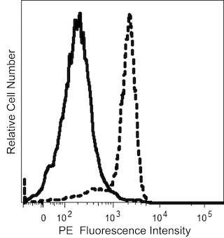
The P-3E10 monoclonal antibody specifically recognizes CD298, the β3 subunit of the Na+/K+ ATPase (Sodium/potassium-transporting ATPase subunit beta-3). CD298 is a ~45-55 kDa single-pass, type II membrane protein that is encoded by ATP1B3 (ATPase Na+/K+ transporting subunit beta 3) which belongs to the P-type ATPases superfamily. CD298 is widely expressed on lymphocytes, monocytes, granulocytes, platelets, and on other hematopoietic and nonhematopoietic cells and cell lines. The Na+/K+ ATPase is an integral membrane protein complex with enzymatic activity that mediates the active transport and exchange of sodium and potassium ions across the plasma membrane. This complex is composed of α and β subunits. The α subunit is a 10-membrane-spanning, catalytic protein that contains binding sites for Na +, K + and ATP. The α subunits are associated with the smaller, regulatory glycoprotein β subunits. Upon ATP hydrolysis, the Na+/K+ ATPase transports Na+ ions out of the cell in exchange for K+ ions that are transported in. This establishes a transmembrane electrochemical gradient that is essential for osmoregulation and for the transport of various nutrients and other molecules by cells. The P-3E10 antibody can reportedly inhibit the activation and proliferation of T cells and B cells.
The antibody was conjugated to BD Horizon™ BUV805 which is part of the BD Horizon Brilliant™ Ultraviolet family of dyes. This dye is a tandem fluorochrome of BD Horizon BUV395 with an Ex Max of 348 nm and an acceptor dye with an Em Max at 805 nm. BD Horizon Brilliant BUV805 can be excited by the ultraviolet laser (355 nm) and detected with a 820/60 filter and a 770LP.

Development References (5)
-
Bouwer AL, Saunderson SC, Caldwell FJ, et al. NK cells are required for dendritic cell-based immunotherapy at the time of tumor challenge.. J Immunol. 2014; 192(5):2514-21. (Clone-specific: Flow cytometry). View Reference
-
P. A. Gorer, S. Lyman, G. D. Snell. Studies on the Genetic and Antigenic Basis of Tumour Transplantation. Linkage between a Histocompatibility Gene and 'Fused' in Mice. Proc R Soc Lond B Biol Sci. 1948; 135(881):499-505. (Biology).
-
Springer TA. Cell-surface differentiation in the mouse. Characterization of "jumping" and "lineage" antigens using xenogeneic rat monoclonal antibodies. In: Kennett RH, McKearn TJ, Bechtol KB, ed. Monoclonal antibodies. Hybridomas: A new dimension in biological analyses. New York and London: Plenum Press; 1980:185-217.
-
Stallcup KC, Springer TA, Mescher MF. Characterization of an anti-H-2 monoclonal antibody and its use in large-scale antigen purification. J Immunol. 1981; 127(30):923-930. (Immunogen: Flow cytometry, Immunoaffinity chromatography, Immunoprecipitation, Radioimmunoassay). View Reference
-
Watanabe Y, Kuribayashi K, Miyatake S, et al. Exogenous expression of mouse interferon gamma cDNA in mouse neuroblastoma C1300 cells results in reduced tumorigenicity by augmented anti-tumor immunity.. Proc Natl Acad Sci USA. 1989; 86(23):9456-60. (Clone-specific: Flow cytometry). View Reference
Please refer to Support Documents for Quality Certificates
Global - Refer to manufacturer's instructions for use and related User Manuals and Technical data sheets before using this products as described
Comparisons, where applicable, are made against older BD Technology, manual methods or are general performance claims. Comparisons are not made against non-BD technologies, unless otherwise noted.
For Research Use Only. Not for use in diagnostic or therapeutic procedures.
Report a Site Issue
This form is intended to help us improve our website experience. For other support, please visit our Contact Us page.