
Preparation And Storage
Recommended Assay Procedures
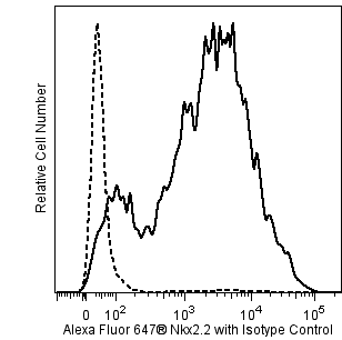
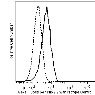
Alexa Fluor® 647 Mouse Anti-Nkx2.2 effectively stains Nkx2.2-transfected 293-F cells when using the BD Pharmingen™ Transcription Factor Buffer Set (Cat. No. 562574). However, we recommend fixation with BD Cytofix™ Fixation Buffer (Cat. No. 554655) and permeabilization with BD Phosflow™ Perm Buffer III (Cat. No. 558050) for staining of untransfected 293-F cells.
Product Notices
- Since applications vary, each investigator should titrate the reagent to obtain optimal results.
- An isotype control should be used at the same concentration as the antibody of interest.
- The Alexa Fluor®, Pacific Blue™, and Cascade Blue® dye antibody conjugates in this product are sold under license from Molecular Probes, Inc. for research use only, excluding use in combination with microarrays, or as analyte specific reagents. The Alexa Fluor® dyes (except for Alexa Fluor® 430), Pacific Blue™ dye, and Cascade Blue® dye are covered by pending and issued patents.
- Alexa Fluor® 647 fluorochrome emission is collected at the same instrument settings as for allophycocyanin (APC).
- Alexa Fluor® is a registered trademark of Molecular Probes, Inc., Eugene, OR.
- Species cross-reactivity detected in product development may not have been confirmed on every format and/or application.
- For fluorochrome spectra and suitable instrument settings, please refer to our Multicolor Flow Cytometry web page at www.bdbiosciences.com/colors.
- Caution: Sodium azide yields highly toxic hydrazoic acid under acidic conditions. Dilute azide compounds in running water before discarding to avoid accumulation of potentially explosive deposits in plumbing.
- Triton is a trademark of the Dow Chemical Company.
- Please refer to www.bdbiosciences.com/us/s/resources for technical protocols.
Companion Products


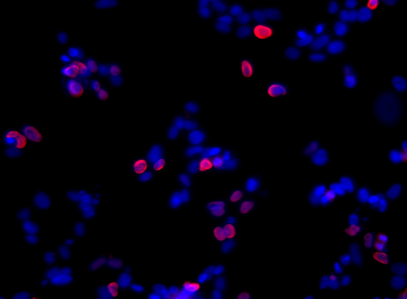
The 74.5A5 monoclonal antibody specifically binds to Homeobox protein Nkx-2.2 (Nkx2.2), which is a transcription factor required for the differentiation of ventral neuronal populations in the central nervous system and endocrine cell populations in the pancreas and intestine. Within the pancreas, Nkx2.2 is expressed in many cell subsets and plays important roles in islet and β-cell differentiation and maturation. Mice with a homozygous disruption of the Nkx2-2 gene lack mature β cells and die shortly after birth with severe hyperglycemia. Similarly to the Nkx2.2-ablated mice, homozygous mutations in the human NKX2-2 gene have been described in diabetic patients and associated with the etiology of neonatal diabetes. The NKX2-2 gene is also a target of the Ewing sarcoma specific protein EWS-FLI. Nkx2.2 expression can be monitored in order to determine cell fate changes in cultures for the generation of human pluripotent stem cell-derived insulin-producing cells as well as oligodendrocyte progenitors.
Development References (6)
-
Briscoe J, Sussel L, Serup P, et al. Homeobox gene Nkx2.2 and specification of neuronal identity by graded Sonic hedgehog signalling.. Nature. 1999; 398(6728):622-7. (Clone-specific: Immunohistochemistry). View Reference
-
D'Amour KA, Bang AG, Eliazer S, et al . Production of pancreatic hormone-expressing endocrine cells from human embryonic stem cells. Nat Biotechnol. 2006; 24(12):1481-1483. (Clone-specific: Immunofluorescence, Western blot). View Reference
-
Ericson J, Rashbass P, Schedl A, et al. Pax6 controls progenitor cell identity and neuronal fate in response to graded Shh signalling. Cell. 1997; 90(1):169-180. (Immunogen: Immunohistochemistry). View Reference
-
Sussel L, Kalamaras J, Hartigan-O'Connor DJ, et al. Mice Lacking the homeodomain transcription factor Nkx2.2 have diabetes due to arrested differentiation of pancreatic beta cells. Development. 1998; 125(12):2213-2221. (Clone-specific: Immunohistochemistry). View Reference
-
Wang S, Bates J, Li X, et al. Human iPSC-derived oligodendrocyte progenitor cells can myelinate and rescue a mouse model of congenital hypomyelination. Cell Stem Cell. 2013; 12(2):252-264. (Clone-specific: Immunofluorescence). View Reference
-
Yoshida A, Sekine S, Tsuta K, Fukayama M, Furuta K, Tsuda H. NKX2.2 is a useful immunohistochemical marker for Ewing sarcoma. Am J Surg Pathol. 2012; 36(7):993-999. (Biology). View Reference
Please refer to Support Documents for Quality Certificates
Global - Refer to manufacturer's instructions for use and related User Manuals and Technical data sheets before using this products as described
Comparisons, where applicable, are made against older BD Technology, manual methods or are general performance claims. Comparisons are not made against non-BD technologies, unless otherwise noted.
For Research Use Only. Not for use in diagnostic or therapeutic procedures.