Dendritic cells – Overview and development
April 01, 2022
Introduction
Dendritic cells (DCs) were originally discovered in 1973 by two Canadian scientists, Ralph Steinmann and Zanvil Cohn as a previously undefined cell type in the mouse spleen. They became known as ‘dendritic cells’ eventually due to their typical features of multiple extended dendritic or pseudopodia-like cytoplasmic protrusions during their maturation stage.1
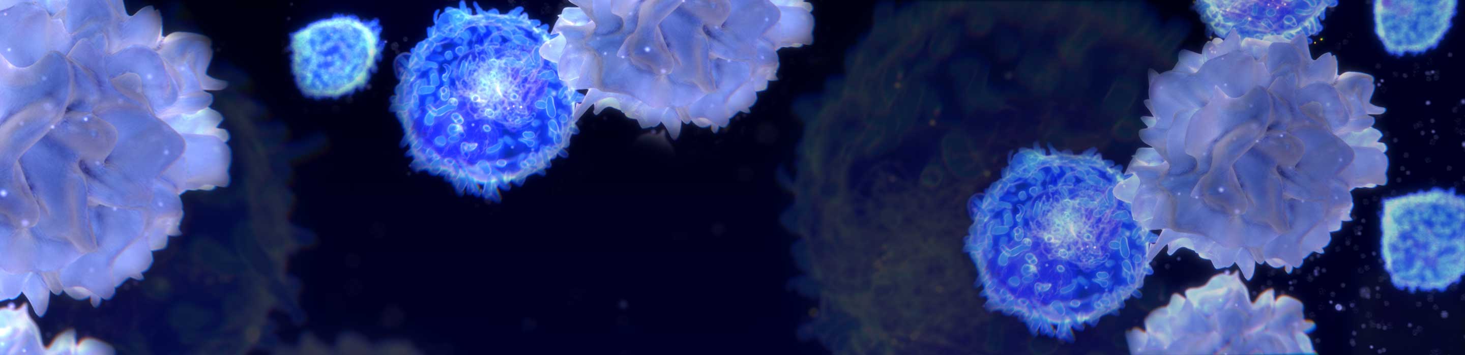
Dendritic cells function as sentinel cells in the immune system and are found in every part of the body including the skin, peripheral blood, mucosal surfaces, interstitial tissues, lymphoid and non-lymphoid tissue areas of the body.1
Origin and anatomical location of dendritic cells
DCs in humans and mice originate from haematopoetic stem cells (HSCs) of the bone marrow, which are derived either from lymphoid or myeloid precursors. The phenotypic features and anatomical functions of these two precursors differ, but both myeloid and lymphoid DCs from humans and mice express a high level of CD11c, MHC-II, and costimulatory molecules CD40, CD80, CD83 and CD86.1 Myeloid and lymphoid DCs can be differentiated based on CD1 markers CD8α and DEC 205.1
Myeloid DCs are primarily located in the spleen marginal zone, while lymphoid DCs are in the T cell areas of the spleen and lymph nodes, precisely in the periarterial lymphatic sheaths (PALS).1
Even though both human and murine DCs originate from the same progenitor, their subsets play different roles in the regulation of B cell activation and the differentiation of T cells into Th-1 and Th-2.
Myeloid precursors express CD14+/CD11C+/CD1- and CD14-/CD11C+/CD1+, whereas the lymphoid precursors express CD14-/CD11C-/IL3Rα. The CD14+/CD11C+/CD1- surface markers expressed by myeloid DCs possess high phagocytic as well as an endocytic ability when compared to lymphoid precursors CD14-/CD11C+/CD1+.1
DCs are the most potent antigen-presenting cells (APCs) that present antigens to T cells. They also play a key role in immunity by inducing innate inflammatory responses to pathogens, priming immature T cells and activating immunological memory T cells. DCs are also involved in other significant immune functions like immune tolerance and adaptive immune response.
Research into DCs has addressed their role in clinical immunology, and their involvement in transplantation, auto-immune disease, cancer and viral infections. They have also been studied as vaccine targets.
The poster
Flow cytometry has contributed immensely to the large bank of information about DC subsets and their markers. The attached poster gives a clear and lucid overview of DC development, including the core DC phenotyping markers, DC subset markers and the secreted cytokines.
Download the “Dendritic cells overview and development poster