





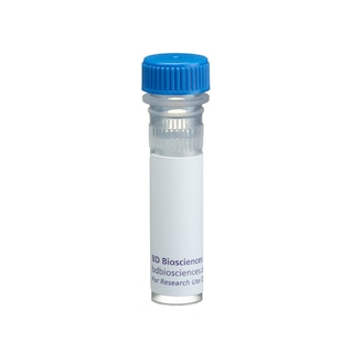
BD Pharmingen™ Purified Rat Anti-Human IL-10
Clone JES3-12G8 (RUO)


Immunohistochemical analysis of human IL-10 expression on human peripheral mononuclear cells. PBMC were isolated from human peripheral blood by density gradient centrifugation and were cultured for 2 days with plate bound anti-human CD3 and soluble anti-human CD28 in the presence of recombinant human IL-2 and recombinant human IL-4. The cells were subsequently harvested, washed and recultured with recombinant human IL-2 and recombinant human IL-4 for an additional 3 days. Finally, the cells were harvested, washed and stimulated with PMA (Sigma) and ionomycin (Sigma) in the presence of GolgiStop™ (Cat. No. 554724) for 4 hours at 37°C. The activated cells were harvested and the presence of IL-10 producing cells was detected by immunocytochemistry using a three-step staining procedure that employs Biotin Goat Anti-Rat Ig (Cat. No. 559286) and Anti-Rat Ig HRP Detection Kit (Cat. No. 551013) (see protocol below) (Nomarski optics, original magnification 400 X). To demonstrate specificity of staining the binding of Purified Rat Anti-Human IL-10 (Cat. No. 559076) antibody was blocked by the preincubation of the purified antibody with excess recombinant human IL-10 protein (Cat. No. 554611; data not shown).

Immunohistochemical analysis of human IL-10 expression on human peripheral mononuclear cells. PBMC were isolated from human peripheral blood by density gradient centrifugation and were cultured for 2 days with plate bound anti-human CD3 and soluble anti-human CD28 in the presence of recombinant human IL-2 and recombinant human IL-4. The cells were subsequently harvested, washed and recultured with recombinant human IL-2 and recombinant human IL-4 for an additional 3 days. Finally, the cells were harvested, washed and stimulated with PMA (Sigma) and ionomycin (Sigma) in the presence of GolgiStop™ (Cat. No. 554724) for 4 hours at 37°C. The activated cells were harvested and the presence of IL-10 producing cells was detected by immunocytochemistry using a three-step staining procedure that employs Biotin Goat Anti-Rat Ig (Cat. No. 559286) and Anti-Rat Ig HRP Detection Kit (Cat. No. 551013) (see protocol below) (Nomarski optics, original magnification 400 X). To demonstrate specificity of staining the binding of Purified Rat Anti-Human IL-10 (Cat. No. 559076) antibody was blocked by the preincubation of the purified antibody with excess recombinant human IL-10 protein (Cat. No. 554611; data not shown).


Preparation And Storage


Recommended Assay Procedures
Immunocytochemistry: Purified Rat Anti-Human IL-10 (Cat. No. 559076) can be used to identify and enumerate human IL-10 producing cells by immunocytochemistry. For optimal indirect immunocytochemical staining, the JES3-12G8 antibody should be titrated and visualized via a three-step staining procedure in combination with biotin goat anti-Rat IgG and an avidin/streptavidin horseradish peroxidase detection system. A detailed protocol for the cytokine immunocytochemical procedure follows.
CYTOKINE IMMUNOCYTOCHEMISTRY PROTOCOL
REAGENTS REQUIRED
1. Fixation Buffer: 5% formalin (10% formalin, CMS, Cat. No. 245-684) is dissolved in phosphate buffered-saline (PBS) (Bacto FA Buffer, Difco Laboratories, Cat. No. 2314-15-0), or BD Pharmingen™ ICC Fixation Buffer (BD Cat. No. 550010)
2. Endogenous Peroxidase Blocking Buffer: DAKO Peroxidase Blocking Reagent (DAKO, Cat. No. S2001).
3. Endogenous Biotin Blocking Buffer: Biotin/Avidin Blocking Kit (Vector Laboratories, Cat. No. SP-2001).
4. Antibody dilution buffer: BD Pharmingen™ Antibody Diluent for IHC, Cat. No. 559148, supplemented with saponin
5. Microscopic slides: Adhesion Slides (Erie Scientific Company, Cat. No. ER-202B-AD) or for cytospins, Colorfrost /Plus slides (Fisher, Cat. No. 12-550-17).
6. Detection system: BD Pharmingen™ Streptavidin-horseradish peroxidase (HRP), (Cat. No. 550946), or Anti-Rat Ig HRP Detection Kit (Cat. No. 551013).
7. Mounting medium for short-term storage: Aqua-mount® (Lerner Laboratories, Cat. No. 13800).
8. DAB Substrate Kit (contains 3-3 -Diaminobenzidine tetra hydrochloride), (BD Cat. No. 550880), or Anti-Rat Ig HRP Detection Kit (Cat. No. 551013).
SECONDARY ANTIBODIES
1. Biotin Goat anti-Rat IgG (Cat. No. 559286) or Anti-Rat Ig HRP Detection Kit (Cat. No. 551013).
PROCEDURE FOR IMMUNOCYTOCHEMICAL STAINING OF SINGLE-CELL PREPARATIONS
This procedure describes the immunoenzymatic technique of staining cytokines within individual cells that are immobilized on microscopic slides via adherence (adherent slides) or centrifugation (cytospins).
ADHESION SLIDES
1. Harvest cells and wash them twice in PBS using centrifugation (400 x g for 5 min) to remove residual protein.
2. Adjust the cell concentration at 4-5 x 10×6 cells/ml in PBS.
3. Place 20 µl of the cell suspension in each well of the adhesion slides and let them adhere at room temperature (RT) for 20 min. Please note that the slides should be washed in PBS at RT for 5 min before transferring the cells.
4. Fix cells on slides using fixation buffer for 15 min at RT.
5. Wash slides 2X in PBS with 5 min incubations.
6. Block slides with PBS supplemented with 1% (w/v) BSA (Sigma, Cat. No. A43-78) for 30 min at RT or 10 min at 37°C.
7. Wash slides 2X in PBS and proceed with staining or air dry them and store them at -80°C for future use.
8. Incubate slides with 20 µl of 1% goat serum and PBS with 0.1% (w/v) saponin for 30 min at RT.
9. Wash slides 2X with PBS with 5 min incubations.
10. Block endogenous peroxidase activity with Endogenous Peroxidase Blocking Buffer (20 µl/well) for 10 min at RT.
11. Wash 2X in PBS with 5 min incubations.
12. Incubate each well with Avidin (20 µl/well) for 15 min.
13. Wash 2X in PBS with 5 min incubations.
14. Incubate each well with Biotin (20 µl/well) for 15 min.
15. Wash 2X in PBS with 5 min incubations.
16. Incubate each well for 1 hr at RT with 20 µl of purified cytokine-specific antibody or appropriate immunoglobulin isotype control diluted in Antibody Diluent for IHC, Cat. No. 559148, supplemented with saponin
17. Wash slides 2X in PBS with 5 min incubations.
18. Incubate each well with 20 µl of a biotinylated secondary antibody diluted in IHC Diluent Buffer for 30 min at RT.
19. Wash 2X in PBS with 5 min incubations.
20. Apply 20 µl of Streptavidin-HRP (BD Cat. No. 550946) to each well on slides and incubate for 30 min at RT.
21. Wash slides 2X with PBS with 5 minutes incubations.
22. Incubate with DAB Substrate as directed, (BD Cat. No. 550880) for less than 5 min at RT.
23. Stop the development of the color reaction by washing with PBS.
24. The slides are subsequently mounted in short-term storage mounting medium.
CYTOSPINS
1. Assemble the Cytospin's sample chamber (e.g. Cytospin 3, Shandon, UK or comparable centrifuge), filter card, slide and cytospin racks according to manufacturer's specifications.
2. Load 40 µl of approximately 1 x 10×6 cells to each sample chamber.
3. Spin slides at 600 rpm for 2 min.
4. Take slides out of the cytospin rack and place them on a staining rack.
5. For fixation and staining please follow the steps 4 through 24 specified above for staining cells on adhesion slides.
Product Notices
- Since applications vary, each investigator should titrate the reagent to obtain optimal results.
- Caution: Sodium azide yields highly toxic hydrazoic acid under acidic conditions. Dilute azide compounds in running water before discarding to avoid accumulation of potentially explosive deposits in plumbing.
- Sodium azide is a reversible inhibitor of oxidative metabolism; therefore, antibody preparations containing this preservative agent must not be used in cell cultures nor injected into animals. Sodium azide may be removed by washing stained cells or plate-bound antibody or dialyzing soluble antibody in sodium azide-free buffer. Since endotoxin may also affect the results of functional studies, we recommend the NA/LE (No Azide/Low Endotoxin) antibody format, if available, for in vitro and in vivo use.
- Species cross-reactivity detected in product development may not have been confirmed on every format and/or application.
- Please refer to http://regdocs.bd.com to access safety data sheets (SDS).
- Please refer to www.bdbiosciences.com/us/s/resources for technical protocols.
Companion Products


The JES3-12G8 antibody reacts with human interleukin-10 (IL-10) and viral IL-10. The immunogen used to generate the JES3-12G8 hybridoma was human IL-10. This is a neutralizing antibody.
Development References (6)
-
Abrams JS, Roncarolo MG, Yssel H, Andersson U, Gleich GJ, Silver JE. Strategies of anti-cytokine monoclonal antibody development: immunoassay of IL-10 and IL-5 in clinical samples. Immunol Rev. 1992; 127:5-24. (Clone-specific: ELISA). View Reference
-
Burdin N, Peronne C, Banchereau J, Rousset F. Epstein-Barr virus transformation induces B lymphocytes to produce human interleukin 10. J Exp Med. 1993; 177(2):295-304. (Clone-specific: ELISA). View Reference
-
Gotlieb WH, Abrams JS, Watson JM, Velu TJ, Berek JS, Martinez-Maza O. Presence of interleukin 10 (IL-10) in the ascites of patients with ovarian and other intra-abdominal cancers. Cytokine. 1992; 4(5):385-390. (Clone-specific: ELISA). View Reference
-
Hsu SM, Raine L, Fanger H. A comparative study of the peroxidase-antiperoxidase method and an avidin-biotin complex method for studying polypeptide hormones with radioimmunoassay antibodies. Am J Clin Pathol. 1981; 75(5):734-738. (Methodology: Immunocytochemistry (cytospins)). View Reference
-
Hsu SM, Raine L, Fanger H. Use of avidin-biotin-peroxidase complex (ABC) in immunoperoxidase techniques: a comparison between ABC and unlabeled antibody (PAP) procedures. J Histochem Cytochem. 1981; 29(4):577-580. (Methodology: Immunocytochemistry (cytospins)). View Reference
-
Yssel H, De Waal Malefyt R, Roncarolo MG, et al. IL-10 is produced by subsets of human CD4+ T cell clones and peripheral blood T cells. J Immunol. 1992; 149(7):2378-2384. (Clone-specific: ELISA). View Reference
Please refer to Support Documents for Quality Certificates
Global - Refer to manufacturer's instructions for use and related User Manuals and Technical data sheets before using this products as described
Comparisons, where applicable, are made against older BD Technology, manual methods or are general performance claims. Comparisons are not made against non-BD technologies, unless otherwise noted.
Please refer to Support Documents for Quality Certificates
Global - Refer to manufacturer's instructions for use and related User Manuals and Technical data sheets before using this products as described
Comparisons, where applicable, are made against older BD Technology, manual methods or are general performance claims. Comparisons are not made against non-BD technologies, unless otherwise noted.
For Research Use Only. Not for use in diagnostic or therapeutic procedures.