-
Reagents
- Flow Cytometry Reagents
-
Western Blotting and Molecular Reagents
- Immunoassay Reagents
-
Single-Cell Multiomics Reagents
- BD® OMICS-Guard Sample Preservation Buffer
- BD® AbSeq Assay
- BD® Single-Cell Multiplexing Kit
- BD Rhapsody™ ATAC-Seq Assays
- BD Rhapsody™ Whole Transcriptome Analysis (WTA) Amplification Kit
- BD Rhapsody™ TCR/BCR Next Multiomic Assays
- BD Rhapsody™ Targeted mRNA Kits
- BD Rhapsody™ Accessory Kits
- BD® OMICS-One Protein Panels
-
Functional Assays
-
Microscopy and Imaging Reagents
-
Cell Preparation and Separation Reagents
-
- BD® OMICS-Guard Sample Preservation Buffer
- BD® AbSeq Assay
- BD® Single-Cell Multiplexing Kit
- BD Rhapsody™ ATAC-Seq Assays
- BD Rhapsody™ Whole Transcriptome Analysis (WTA) Amplification Kit
- BD Rhapsody™ TCR/BCR Next Multiomic Assays
- BD Rhapsody™ Targeted mRNA Kits
- BD Rhapsody™ Accessory Kits
- BD® OMICS-One Protein Panels
- Luxembourg (English)
-
Change country/language
Old Browser
This page has been recently translated and is available in French now.
Looks like you're visiting us from United States.
Would you like to stay on the current country site or be switched to your country?
BD Pharmingen™ Purified NA/LE Mouse Anti-Human CD3
Clone UCHT1 (also known as UCHT-1; UCHT 1) (RUO)

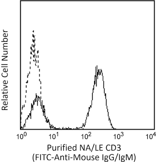
Flow cytometric analysis of CD3 expression on human peripheral blood lymphocytes. Human whole blood was stained with either Purified NA/LE Mouse IgG1 κ Isotype Control (Cat. No. 554721; dashed line histogram) or Purified NA/LE Mouse Anti-Human CD3 antibody (Cat. No. 555329/562280/562310; solid line histogram). Erythrocytes were lysed with BD Pharm Lyse™ Lysing Buffer (Cat. No. 555899). The cells were washed and then stained with FITC Goat Anti-Mouse IgG/IgM (Cat. No. 562292; dashed line histogram). The erythrocytes were lysed with BD Pharm Lyse™ Lysing Buffer (Cat. No. 555899). The fluorescence histograms were derived from events with the forward and side light-scatter characteristics of viable lymphocytes. Flow cytometry was performed using a BD™ LSR II Flow Cytometer System.


Flow cytometric analysis of CD3 expression on human peripheral blood lymphocytes. Human whole blood was stained with either Purified NA/LE Mouse IgG1 κ Isotype Control (Cat. No. 554721; dashed line histogram) or Purified NA/LE Mouse Anti-Human CD3 antibody (Cat. No. 555329/562280/562310; solid line histogram). Erythrocytes were lysed with BD Pharm Lyse™ Lysing Buffer (Cat. No. 555899). The cells were washed and then stained with FITC Goat Anti-Mouse IgG/IgM (Cat. No. 562292; dashed line histogram). The erythrocytes were lysed with BD Pharm Lyse™ Lysing Buffer (Cat. No. 555899). The fluorescence histograms were derived from events with the forward and side light-scatter characteristics of viable lymphocytes. Flow cytometry was performed using a BD™ LSR II Flow Cytometer System.

Flow cytometric analysis of CD3 expression on human peripheral blood lymphocytes. Human whole blood was stained with either Purified NA/LE Mouse IgG1 κ Isotype Control (Cat. No. 554721; dashed line histogram) or Purified NA/LE Mouse Anti-Human CD3 antibody (Cat. No. 555329/562280/562310; solid line histogram). Erythrocytes were lysed with BD Pharm Lyse™ Lysing Buffer (Cat. No. 555899). The cells were washed and then stained with FITC Goat Anti-Mouse IgG/IgM (Cat. No. 562292; dashed line histogram). The erythrocytes were lysed with BD Pharm Lyse™ Lysing Buffer (Cat. No. 555899). The fluorescence histograms were derived from events with the forward and side light-scatter characteristics of viable lymphocytes. Flow cytometry was performed using a BD™ LSR II Flow Cytometer System.


BD Pharmingen™ Purified NA/LE Mouse Anti-Human CD3

Regulatory Status Legend
Any use of products other than the permitted use without the express written authorization of Becton, Dickinson and Company is strictly prohibited.
Preparation And Storage
Product Notices
- Since applications vary, each investigator should titrate the reagent to obtain optimal results.
- An isotype control should be used at the same concentration as the antibody of interest.
- Please refer to http://regdocs.bd.com to access safety data sheets (SDS).
- Please refer to www.bdbiosciences.com/us/s/resources for technical protocols.
Data Sheets
Companion Products


The UCHT1 monoclonal antibody specifically binds to the human CD3ε-chain, a 20-kDa subunit of the CD3/T cell antigen receptor complex. CD3ε is expressed on 70-80% of normal human peripheral blood lymphocytes and 60-85% of thymocytes. Studies from the HLDA Workshop show that this antibody is mitogenic for CD3ε-positive cells when used in conjunction with costimulatory agents such as pokeweed mitogen or anti-CD28 antibody. CD3 plays a central role in signal transduction during antigen recognition. The UCHT1 antibody stains both surface and intracellular CD3ε unlike the other CD3 clone, HIT3a, that stains only extracellular CD3ε.
Development References (9)
-
Bernard A, Boumsell L, Dausset J, Milstein C, Schlossman SF, ed. Leukocyte Typing. New York: Springer-Verlag; 1984:1-814.
-
Beverley PC, Callard RE. Distinctive functional characteristics of human "T" lymphocytes defined by E rosetting or a monoclonal anti-T cell antibody. Eur J Immunol. 1981; 11(4):329-334. (Clone-specific). View Reference
-
Burns GF, Boyd AW, Beverley PC. Two monoclonal anti-human T lymphocyte antibodies have similar biologic effects and recognize the same cell surface antigen. J Immunol. 1982; 129(4):1451-1457. (Clone-specific: Blocking, Functional assay, Inhibition). View Reference
-
Knapp W. W. Knapp .. et al., ed. Leucocyte typing IV : white cell differentiation antigens. Oxford New York: Oxford University Press; 1989:1-1182.
-
Lanier LL, Allison JP, Phillips JH. Correlation of cell surface antigen expression on human thymocytes by multi-color flow cytometric analysis: implications for differentiation. J Immunol. 1986; 137(8):2501-2507. (Biology). View Reference
-
McMichael AJ. A.J. McMichael .. et al., ed. Leucocyte typing III : white cell differentiation antigens. Oxford New York: Oxford University Press; 1987:1-1050.
-
Schlossman SF. Stuart F. Schlossman .. et al., ed. Leucocyte typing V : white cell differentiation antigens : proceedings of the fifth international workshop and conference held in Boston, USA, 3-7 November, 1993. Oxford: Oxford University Press; 1995.
-
Van Wauwe JP, Goossens JG, Beverley PC. Human T lymphocyte activation by monoclonal antibodies; OKT3, but not UCHT1, triggers mitogenesis via an interleukin 2-dependent mechanism. J Immunol. 1984; 133(1):129-132. (Clone-specific: Functional assay). View Reference
-
Zola H. Leukocyte and stromal cell molecules : the CD markers. Hoboken, N.J.: Wiley-Liss; 2007.
Please refer to Support Documents for Quality Certificates
Global - Refer to manufacturer's instructions for use and related User Manuals and Technical data sheets before using this products as described
Comparisons, where applicable, are made against older BD Technology, manual methods or are general performance claims. Comparisons are not made against non-BD technologies, unless otherwise noted.
For Research Use Only. Not for use in diagnostic or therapeutic procedures.